Human VE-Cadherin Antibody
R&D Systems, part of Bio-Techne | Catalog # MAB9381


Key Product Details
Validated by
Species Reactivity
Validated:
Cited:
Applications
Validated:
Cited:
Label
Antibody Source
Product Specifications
Immunogen
Asp48-Gln593
Accession # P33151
Specificity
Clonality
Host
Isotype
Scientific Data Images for Human VE-Cadherin Antibody
Detection of Human VE‑Cadherin by Western Blot.
Western blot shows lysate of HUVEC human umbilical vein endothelial cells. PVDF membrane was probed with 1 µg/mL of Mouse Anti-Human VE-Cadherin Monoclonal Antibody (Catalog # MAB9381) followed by HRP-conjugated Anti-Mouse IgG Secondary Antibody (Catalog # HAF018). A specific band was detected for VE-Cadherin at approximately 125 kDa (as indicated). This experiment was conducted under reducing conditions and using Immunoblot Buffer Group 1.VE‑Cadherin in HUVEC Cells.
VE-Cadherin was detected in immersion fixed HUVEC cells using Mouse Anti-Human VE-Cadherin Monoclonal Antibody (Catalog # MAB9381) at 0.5 µg/mL for 3 hours at room temperature. Cells were stained using the NorthernLights™ 557-conjugated Anti-Mouse IgG Secondary Antibody (red; Catalog # NL007) and counterstained with DAPI (blue). Specific staining was localized to plasma membrane. View our protocol for Fluorescent ICC Staining of Cells on Coverslips.Detection of Human VE-Cadherin by Flow Cytometry
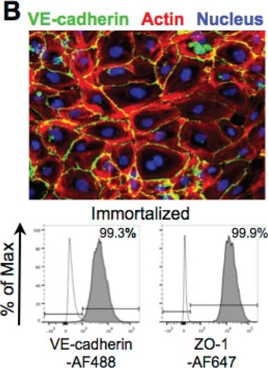
Immortalized HAECs retain phenotypic and functional characteristics of primary cells. (A) (Top) Immortalized HAEC confluent monolayers stained with CD31-AF488 antibody (green) and DAPI (blue). (Bottom) Flow cytometry histogram plots of primary or immortalized HAECs labeled with CD31-AF488 antibody. Secondary antibody only (dashed line) and nonstained cells (dotted line) were used as negative controls. (B) (Top) Immortalized HAEC confluent monolayers stained with VE-cadherin-AF488 (green) antibody, Acti-Stain 555 phalloidin (red), and DAPI (blue). (Bottom) Flow cytometry histogram plots of immortalized HAECs labeled with VE-cadherin-AF488 and ZO-1-AF647 antibodies. Secondary antibody only (dashed line) was used as a negative control. (C) Tube forming assay of primary (top) and immortalized (bottom) HAECs at 0 h and 8 h after seeding in Matrigel-coated wells. (D) Acetylated low-density lipoprotein (Ac-LDL) uptake assay of primary HAECs (top), immortalized HAECs (middle), and fibroblasts (negative control; bottom) treated with fluorescently labeled Ac-LDL (red) for 2.5 h and stained with DAPI (blue). (E) IL-8 detection in culture supernatants 24 h after treatment of primary or immortalized HAECs with increasing concentrations of TNF-alpha, IFN-gamma, or IL-6. (F) IL-8 detection in culture supernatants 24 h after treatment of immortalized HAECs with increasing concentrations of IL-2 or IL-1. Image collected and cropped by CiteAb from the following publication (https://pubmed.ncbi.nlm.nih.gov/29229737), licensed under a CC-BY license. Not internally tested by R&D Systems.Applications for Human VE-Cadherin Antibody
CyTOF-ready
Flow Cytometry
Sample: HUVEC human umbilical vein endothelial cells stained in buffer containing Ca2+ and Mg2+
Immunocytochemistry
Sample: Immersion fixed HUVEC human umbilical vein endothelial cells
Western Blot
Sample: HUVEC human umbilical vein endothelial cells
Reviewed Applications
Read 2 reviews rated 4.5 using MAB9381 in the following applications:
Formulation, Preparation, and Storage
Purification
Reconstitution
Formulation
Shipping
Stability & Storage
- 12 months from date of receipt, -20 to -70 °C as supplied.
- 1 month, 2 to 8 °C under sterile conditions after reconstitution.
- 6 months, -20 to -70 °C under sterile conditions after reconstitution.
Background: VE-Cadherin
Vascular endothelial (VE)-cadherin (VE-CAD), also called 7B4 and cadherin‑5 (CDH5), is a member of the cadherin family of cell adhesion molecules. Cadherins are calcium‑dependent transmembrane proteins which bind to one another in a homophilic manner. On their cytoplasmic side, they associate with the three catenins, alpha, beta, and gamma (plakoglobin). This association links the cadherin protein to the cytoskeleton. Without association with the catenins, the cadherins are non-adhesive. Cadherins play a role in development, specifically in tissue formation. They may also help to maintain tissue architecture in the adult. VE-cadherin has been shown to play important roles in vasculogenesis and angiogenesis. VE-cadherin is a classical cadherin molecule. Classical cadherins consist of a large extracellular domain which contains DXD and DXNDN repeats responsible for mediating calcium-dependent adhesion, a single-pass transmembrane domain, and a short carboxy-terminal cytoplasmic domain responsible for interacting with the catenins. Human VE-cadherin is a 784 amino acid (aa) residue protein with a 25 aa signal sequence and a 759 aa propeptide. The mature protein begins at amino acid 48 and has a 546 aa extracellular domain, a 27 aa transmembrane domain, and a 164 aa cytoplasmic domain. The human and mouse mature VE-cadherin proteins share approximately 74% homology.
References
- Shimoyama, Y. et al. (1989) J. Cell Biol. 109:1787.
- Bussemakers, M.J.G. et al. (1993) Mol. Biol. Reports 17:123.
- Overduin, M. et al. (1995) Science 267:386.
- Takeichi, M. (1991) Science 251:1451.
- Nose, A. et al. (1987) EMBO J. 6:3655.
- Carmeliet, P. et al. (1999) Cell 98:147.
- Gory-Faure, S. et al. (1999) Development 126:2093.
Long Name
Alternate Names
Gene Symbol
UniProt
Additional VE-Cadherin Products
Product Documents for Human VE-Cadherin Antibody
Product Specific Notices for Human VE-Cadherin Antibody
For research use only

