IL-1 beta/IL-1F2 Antibody - Azide and BSA Free
Novus Biologicals, part of Bio-Techne | Catalog # NB600-633

Key Product Details
Validated by
Biological Validation
Species Reactivity
Validated:
Human, Mouse, Rat, Porcine, Canine, Golden Syrian Hamster, Monkey
Cited:
Human, Mouse, Rat, Porcine, Bacteria, Canine, Golden Syrian Hamster, Ovine, Primate - Macaca mulatta (Rhesus Macaque)
Applications
Validated:
Dot Blot, Electron Microscopy, ELISA, Flow Cytometry, Immunocytochemistry/ Immunofluorescence, Immunohistochemistry, Immunohistochemistry Whole-Mount, Immunohistochemistry-Frozen, Immunohistochemistry-Paraffin, Immunoprecipitation, Western Blot
Cited:
Block/Neutralize, Electron Microscopy, ELISA, IF/IHC, Immunocytochemistry/ Immunofluorescence, Immunohistochemistry, Immunohistochemistry Whole-Mount, Immunohistochemistry-Frozen, Immunohistochemistry-Paraffin, Immunoprecipitation, Western Blot
Label
Unconjugated
Antibody Source
Polyclonal Rabbit IgG
Format
Azide and BSA Free
Concentration
Please see the vial label for concentration. If unlisted please contact technical services.
Product Specifications
Immunogen
This IL-1 beta/IL-1F2 Antibody was prepared by repeated immunizations with recombinant human IL-1 beta/IL-1F2 produced in E.coli. The MW of the recombinant 153 aa IL-1 beta/IL-1F2 was 17 kDa with the N-terminal amino acid at position alanine 117. This cleavage site is generated by the IL-1 beta/IL-1F2 converting enzyme (ICE, capase-1). (Uniprot: P01584)
Reactivity Notes
In general, this antibody also detects primate IL-1 beta/IL-1F2 in the same formats using similar dilutions. Use in Mouse reported in scientific literature (PMID: 33731931).
Localization
Secreted
Specificity
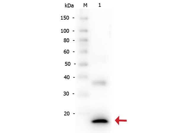
This antibody is primarily directed against mature, 17,000 MW human IL-1 beta/IL-1F2and is useful in determining its presence in various assays. In general, this antibody also detects primate IL-1 beta/IL-1F2in the same formats using similar dilutions. The antiserum does not recognize human IL-1alpha. In ELISA formats and other immunoreactive assays, this antibody will recognize 10% of the non-denatured (native) precursor 31,000 MW IL-1 beta/IL-1F2containing samples but will primarily detect all of the 17,000 MW mature molecule. However, in immunoblot analysis of natural cell products or human body fluids, the usual procedure of hearing the sample in SDS with or without reducing agents will facilitate denaturing of the 31,000 MW IL- 1beta precursor molecule. Denatured 31,000 precursor IL-1 beta/IL-1F2will be recognized by this antibody but often migrates as a 35,000 MW band. This is due to the unfolding of the denatured precursor IL-1 beta/IL-1F2exposing epitopes not exposed in the natural state. In immunoblots, depending on the number of cells, the antibody detects the 17,000 MW band in supernatants as well as a 35,000 MW band representing the 31,000 MW IL-1 beta/IL-1F2precursor in lysates.
Clonality
Polyclonal
Host
Rabbit
Isotype
IgG
Description
This is an IgG preparation of whole rabbit serum purified by DEAE fractionation. Store vial at -20C prior to opening. Aliquot contents and freeze at -20C or below for extended storage. Avoid cycles of freezing and thawing. Centrifuge product if not completely clear after standing at room temperature. This product is stable for several weeks at 4C as an undiluted liquid. Dilute only prior to immediate use.
Scientific Data Images for IL-1 beta/IL-1F2 Antibody - Azide and BSA Free
Western Blot: IL-1 beta/IL-1F2 Antibody [NB600-633]
Western Blot: IL-1 beta/IL-1F2 Antibody [NB600-633] - Methylene blue (M) inhibits NLRP3 inflammasome formation in post-spinal cord injury (SCI) microglia compared to sham (V). Expression of pro-IL-1B and mature IL-1B in microglia sorted from post-SCI spinal cords. Representative Western blot images are shown in (B). Image collected and cropped by CiteAb from the following publication (https://journal.frontiersin.org/article/10.3389/fncel.2017.00391/full) licensed under a CC-BY license.Immunocytochemistry/ Immunofluorescence: IL-1 beta/IL-1F2 Antibody [NB600-633]
Immunocytochemistry/Immunofluorescence: IL-1 beta/IL-1F2 Antibody [NB600-633] - Trabecular Meshwork (TM) region of pig eyes were stained with IL-1 beta antibody (red). Image from verified customer review.Immunohistochemistry: IL-1 beta/IL-1F2 Antibody [NB600-633]
Immunohistochemistry: IL-1 beta/IL-1F2 Antibody [NB600-633] - Analysis of: Human IL1beta antibody Secondary antibody: Peroxidase goat anti-rabbit at 1:10,000 for 45 min at RT Localization: cytoplasm Staining: Close up of medullary lymph node: positive staining in the cytoplasm of circulating macrophages. Neg Ctr (far right) normal rabbit IgG with pH 6.2 40XApplications for IL-1 beta/IL-1F2 Antibody - Azide and BSA Free
Application
Recommended Usage
ELISA
1:500-1:2000
Electron Microscopy
1:10-1:500
Immunocytochemistry/ Immunofluorescence
1:10-1:500
Immunohistochemistry
1:100-1:200
Immunohistochemistry-Paraffin
1:10-1:500
Immunoprecipitation
1:400-1:800
Western Blot
1:1000
Application Notes
This product has been tested for use in ELISA, immunohistochemistry, immunoblotting. This antibody is suitable for neutralizations, radioimmunoassays, flow cytometry, and immunoprecipitation. It recognizes the 17,000 MW mature IL-1beta. For immunoblots, typically, IL-1beta is detected from supernatants or lysates of 2 x 10E6 endotoxin-stimulated peripheral blood mononuclear cells (PBMC). PBMC are stimulated for 24 hours with 1% (v/v) serum plus 10 ng/mL E.coli LPS. For immunoprecipitation pre-clearing the preparation with a non-specific Rabbit IgG to reduce background is suggested. For immunohistochemistry either paraffin fixation or cryofixation can be used for sample preparation to stain intracellular IL-1beta. For ELISA use HRP Conjugated Anti-Rabbit IgG [H&L] (Goat) (611-1302) for detection. In ELISA formats this antibody is best used as the second antibody in combination with a monoclonal antibody as a capture antibody. This antibody is also useful for neutralization of human and primate IL-1beta activity in bioassays. It does not neutralize the biological activity IL-1alpha. It does not neutralize the biological activity of murine, rat or rabbit IL-1beta. For neutralization, it is recommended to incubate the sample with a dilution of the antibody for at least 4 hours before being tested. A control of similarly diluted normal rabbit IgG is recommended. This antibody can be used for FACS analysis. Caution should be exhibited as the F(c) domain of the rabbit IgG molecule may interact with cells non-specifically.
Use in Immunohistochemistry-Frozen reported in scientific literature (PMID: 22898394).
Use in Immunohistochemistry Whole-Mount reported in scientific literature (PMID:31399621).
Use in Immunohistochemistry-Frozen reported in scientific literature (PMID: 22898394).
Use in Immunohistochemistry Whole-Mount reported in scientific literature (PMID:31399621).
Reviewed Applications
Read 5 reviews rated 4 using NB600-633 in the following applications:
Formulation, Preparation, and Storage
Purification
Ion exchange chromatography
Formulation
0.02 M Potassium Phosphate, 0.15 M Sodium Chloride, pH 7.2
Format
Azide and BSA Free
Preservative
No Preservative
Concentration
Please see the vial label for concentration. If unlisted please contact technical services.
Shipping
The product is shipped with polar packs. Upon receipt, store it immediately at the temperature recommended below.
Stability & Storage
Store at -20C short term. Aliquot and store at -80C long term. Avoid freeze-thaw cycles.
Background: IL-1 beta/IL-1F2
IL-1 beta binding to its receptor IL-1RI and the downstream signaling contributes to a dual pathophysiological role (3). On one hand, IL-1 beta signaling activates immune cells and drives CD4+ T cell polarization to T helper type 1 (Th1) and Th17 cells, resulting in anti-tumor responses and mediation of acute inflammation (2,3). However, IL-1 beta also supports tumor growth and metastasis driven by multiple mechanisms including chronic inflammation, an immunosuppressive tumor microenvironment (TME), and angiogenesis (3). Additionally, IL-1 beta signaling been implicated in the pathogenesis of neuroinflammatory diseases of the central nervous system (CNS) such as multiple sclerosis (MS), Alzheimer's disease, and diabetic retinopathy (DR) (2). Mouse studies have shown regression of tumors treated with IL-1 as well as protective effects of IL-1 beta in instances of induced colitis and colon carcinoma (3). Conversely, blocking IL-1 beta has also shown promising effect in cancer treatment, especially when combined with chemotherapeutics (2,3). Approved IL-1 beta monoclonal antibody canakinumab has shown significant therapeutic promise in the treatment of DR (2). Given its multifaceted role in disease, IL-1 beta is a promising therapeutic target.
References
1. Lopez-Castejon G, Brough D. Understanding the mechanism of IL-1beta secretion. Cytokine Growth Factor Rev. 2011;22(4):189-195. https://doi.org/10.1016/j.cytogfr.2011.10.001
2. Mendiola AS, Cardona AE. The IL-1beta phenomena in neuroinflammatory diseases. J Neural Transm (Vienna). 2018;125(5):781-795. https://doi.org/10.1007/s00702-017-1732-9
3. Bent R, Moll L, Grabbe S, Bros M. Interleukin-1 Beta-A Friend or Foe in Malignancies?. Int J Mol Sci. 2018;19(8):2155. https://doi.org/doi:10.3390/ijms19082155
4. Krumm B, Xiang Y, Deng J. Structural biology of the IL-1 superfamily: key cytokines in the regulation of immune and inflammatory responses. Protein Sci. 2014;23(5):526-538. https://doi.org/10.1002/pro.2441
5. He Y, Hara H, Nunez G. Mechanism and Regulation of NLRP3 Inflammasome Activation. Trends Biochem Sci. 2016;41(12):1012-1021. https://doi.org/10.1016/j.tibs.2016.09.002
6. Uniprot (P01584)
Long Name
Interleukin 1 beta
Alternate Names
IL-1b, IL-1F2, IL1 beta, IL1B
Gene Symbol
IL1B
UniProt
Additional IL-1 beta/IL-1F2 Products
Product Documents for IL-1 beta/IL-1F2 Antibody - Azide and BSA Free
Product Specific Notices for IL-1 beta/IL-1F2 Antibody - Azide and BSA Free
This product is for research use only and is not approved for use in humans or in clinical diagnosis. Primary Antibodies are guaranteed for 1 year from date of receipt.
Loading...
Loading...
Loading...
Loading...
Loading...
![Immunocytochemistry/ Immunofluorescence: IL-1 beta/IL-1F2 Antibody [NB600-633] Immunocytochemistry/ Immunofluorescence: IL-1 beta/IL-1F2 Antibody [NB600-633]](https://resources.bio-techne.com/images/products/IL-1-beta-IL-1F2-Antibody-Immunocytochemistry-Immunofluorescence-NB600-633-img0009.jpg)
![Immunohistochemistry: IL-1 beta/IL-1F2 Antibody [NB600-633] Immunohistochemistry: IL-1 beta/IL-1F2 Antibody [NB600-633]](https://resources.bio-techne.com/images/products/IL-1-beta-IL-1F2-Antibody-Immunohistochemistry-NB600-633-img0010.jpg)
![Western Blot: IL-1 beta/IL-1F2 Antibody [NB600-633] Western Blot: IL-1 beta/IL-1F2 Antibody [NB600-633]](https://resources.bio-techne.com/images/products/IL-1-beta-IL-1F2-Antibody-Western-Blot-NB600-633-img0005.jpg)
![Western Blot: IL-1 beta/IL-1F2 Antibody [NB600-633] Western Blot: IL-1 beta/IL-1F2 Antibody [NB600-633]](https://resources.bio-techne.com/images/products/IL-1-beta-IL-1F2-Antibody-Western-Blot-NB600-633-img0012.jpg)
![Western Blot: IL-1 beta/IL-1F2 Antibody [NB600-633] Western Blot: IL-1 beta/IL-1F2 Antibody [NB600-633]](https://resources.bio-techne.com/images/products/IL-1-beta-IL-1F2-Antibody-Western-Blot-NB600-633-img0006.jpg)
![Western Blot: IL-1 beta/IL-1F2 Antibody [NB600-633] Western Blot: IL-1 beta/IL-1F2 Antibody [NB600-633]](https://resources.bio-techne.com/images/products/IL-1-beta-IL-1F2-Antibody-Western-Blot-NB600-633-img0008.jpg)
![Western Blot: IL-1 beta/IL-1F2 Antibody [NB600-633] Western Blot: IL-1 beta/IL-1F2 Antibody [NB600-633]](https://resources.bio-techne.com/images/products/IL-1-beta-IL-1F2-Antibody-Western-Blot-NB600-633-img0011.jpg)

![Western Blot: IL-1 beta/IL-1F2 Antibody [NB600-633] - IL-1 beta/IL-1F2 Antibody](https://resources.bio-techne.com/images/products/nb600-633_rabbit-polyclonal-il-1-beta-il-1f2-antibody-310202416165633.jpg)
![Western Blot: Rabbit Polyclonal IL-1 beta/IL-1F2 Antibody [NB600-633] IL-1 beta/IL-1F2 Antibody](https://resources.bio-techne.com/images/products/antibody/nb600-633_rabbit-polyclonal-il-1-beta-il-1f2-antibody-western-blot-25112024153834..png)