Mouse CCR3 Fluorescein-conjugated Antibody
R&D Systems, part of Bio-Techne | Catalog # FAB729F


Conjugate
Catalog #
Key Product Details
Species Reactivity
Validated:
Mouse
Cited:
Mouse
Applications
Validated:
Flow Cytometry
Cited:
Flow Cytometry, Immunocytochemistry, Immunohistochemistry
Label
Fluorescein (Excitation = 488 nm, Emission = 515-545 nm)
Antibody Source
Monoclonal Rat IgG2A Clone # 83101
Product Specifications
Immunogen
Y3 rat myeloid cell line transfected with mouse CCR3
Met1-Phe359
Accession # P51678
Met1-Phe359
Accession # P51678
Specificity
Detects mouse CCR3. Specifically
stains mouse CCR3 transfectants but not the parental cell lines. Does
not cross‑react with Y3 cells transfected with human CCR3.
Clonality
Monoclonal
Host
Rat
Isotype
IgG2A
Scientific Data Images for Mouse CCR3 Fluorescein-conjugated Antibody
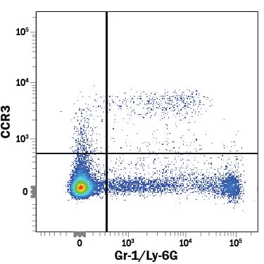
Detection of CCR3 in Mouse Splenocytes by Flow Cytometry.
Mouse splenocytes were stained with Rat Anti-Mouse CCR3 Fluorescein-conjugated Monoclonal Antibody (Catalog # FAB729F) and Rat Anti-Mouse Gr-1/Ly-6G APC-conjugated Monoclonal Antibody (Catalog # FAB1037A). Quadrant markers were set based on control antibody staining (Catalog # IC006F). View our protocol for Staining Membrane-associated Proteins.Detection of Mouse CCR3 by Flow Cytometry
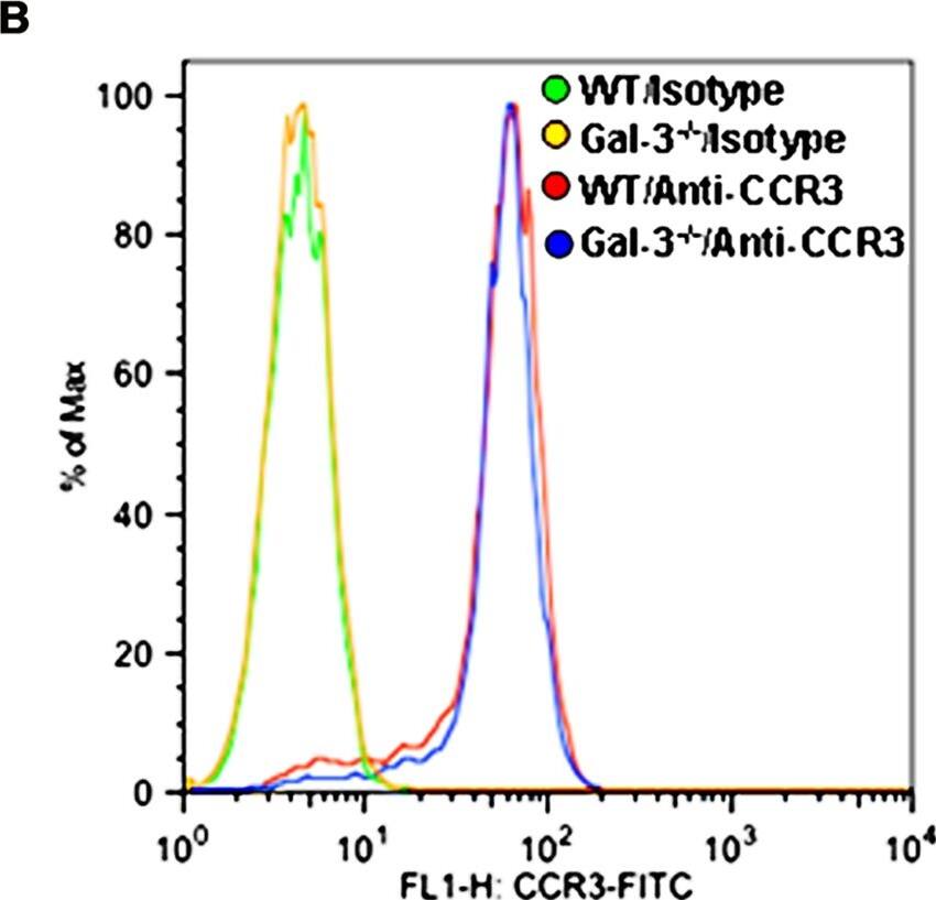
Gal-3 deficient Eos exhibit decreased migration toward eotaxin-1. (A) Migration of WT and Gal-3-/- BM-derived Eos toward murine eotaxin-1 (100 nM) in vitro using 96-well Transwell ® Chambers. Results are represented as percent cell migration relative to WT Eos. (B) Expression of CCR3 by WT and Gal-3-/- Eos by flow cytometry using FITC-conjugated rat anti-mouse CCR3 with FITC-conjugated rat IgG2a as isotype control. Representative data of two independent experiments with Eos from different mice is shown. (C) Migration of WT Eos suspended in medium alone or medium containing lactose or maltose toward murine eotaxin-1. Number of cells that migrated in each case was determined and expressed as the average number of cells/field. Combined data (mean ± SEM) from three independent experiments in duplicate is shown in (A) and (C). #p <0.05 versus WT Eos in (A). Image collected and cropped by CiteAb from the following publication (https://journal.frontiersin.org/article/10.3389/fphar.2013.00037/abstract), licensed under a CC-BY license. Not internally tested by R&D Systems.Applications for Mouse CCR3 Fluorescein-conjugated Antibody
Application
Recommended Usage
Flow Cytometry
10 µL/106 cells
Sample: Mouse splenocytes
Sample: Mouse splenocytes
Formulation, Preparation, and Storage
Purification
Protein A or G purified from hybridoma culture supernatant
Formulation
Supplied in a saline solution containing BSA and Sodium Azide.
Shipping
The product is shipped with polar packs. Upon receipt, store it immediately at the temperature recommended below.
Stability & Storage
Protect from light. Do not freeze.
- 12 months from date of receipt, 2 to 8 °C as supplied.
Background: CCR3
Alternate Names
CCR3, CD193
Gene Symbol
CCR3
UniProt
Additional CCR3 Products
Product Documents for Mouse CCR3 Fluorescein-conjugated Antibody
Product Specific Notices for Mouse CCR3 Fluorescein-conjugated Antibody
For research use only
Loading...
Loading...
Loading...
Loading...
Loading...
