Mouse CX3CL1/Fractalkine Chemokine Domain Antibody
R&D Systems, part of Bio-Techne | Catalog # MAB571


Conjugate
Catalog #
Key Product Details
Validated by
Biological Validation
Species Reactivity
Validated:
Mouse
Cited:
Mouse
Applications
Validated:
ELISA Capture (Matched Antibody Pair), Neutralization, Western Blot
Cited:
Immunocytochemistry, Immunohistochemistry, Immunohistochemistry-Frozen, In vivo assay, Neutralization
Label
Unconjugated
Antibody Source
Monoclonal Rat IgG2A Clone # 126315
Product Specifications
Immunogen
Mouse myeloma cell line NS0-derived recombinant mouse CX3CL1/Fractalkine, S. frugiperda insect ovarian cell line Sf 21-derived recombinant mouse CX3CL1/Fractalkine, and E. coli-derived recombinant mouse CX3CL1/Fractalkine
Specificity
Detects mouse CX3CL1/Fractalkine in Western blots. In direct ELISAs, this antibody shows 100% cross-reactivity with the chemokine domain of recombinant human (rh) CX3CL1, 13% cross-reactivity with rhCX3CL1, and 50% cross-reactivity with recombinant rat CX3CL1.
Clonality
Monoclonal
Host
Rat
Isotype
IgG2A
Endotoxin Level
<0.10 EU per 1 μg of the antibody by the LAL method.
Scientific Data Images for Mouse CX3CL1/Fractalkine Chemokine Domain Antibody
Chemotaxis Induced by CX3CL1/Fractalkine and Neutralization by Mouse CX3CL1/Fractalkine Antibody.
Recombinant Mouse CX3CL1/Fractalkine (Catalog # 571-MF) chemoattracts the BaF3 mouse pro-B cell line transfected with human CX3CR1 in a dose-dependent manner (orange line). The amount of cells that migrated through to the lower chemotaxis chamber was measured by Resazurin (Catalog # AR002). Chemotaxis elicited by Recombinant Mouse CX3CL1/Fractalkine (30 ng/mL) is neutralized (green line) by increasing concentrations of Rat Anti-Mouse CX3CL1/Fractalkine Chemokine Domain Monoclonal Antibody (Catalog # MAB571). The ND50 is typically 0.75-3.5 µg/mL.Detection of Mouse CX3CL1/Fractalkine by Immunocytochemistry/Immunofluorescence
CX3CL1 expression in the brain during EAE progression. (A) Brain CX3CL1 expression of wild type mice with EAE as determined by quantitative real-time PCR. Gene levels were normalized to GAPDH levels and are displayed as levels relative to naïve mice. Error bars represent the s.e.m. (n = 5 mice per group). (B-M) Brains from naïve wild type mice were harvested and frozen for immunostaining. CX3CL1 expression (red) and DAPI nuclei staining (blue) in (B, F, J) naïve and (C, G, K) Day 10, (D, H, L) Day 14, and (E, I, M) Day 21 post-EAE induced wild type mice. CX3CL1 expression is displayed at the (B-E) choroid plexus, (F-I) in and near the hippocampus, and (J-M) cerebellum. White scale bars represent 50 μm. (N) CX3CL1 expression (relative to non-treated cells) in the CPLacZ-2 mouse choroid plexus cell line after 2 hour treatment with varying concentrations of the A2A adenosine receptor specific adenosine receptor agonist CGS21680. Error bars represent the s.e.m. (O) Lymphocyte migration across a transwell choroid plexus barrier following pretreatment with vehicle treatment alone, CGS21680, or CGS21680 and anti-CX3CL1. Total migration was normalized to the vehicle control (set to 100%). Error bars represent the s.e.m. These results are representative of two separate experiments (n ≤ 3). Image collected and cropped by CiteAb from the following publication (https://jneuroinflammation.biomedcentral.com/articles/10.1186/1742-2094-9-193), licensed under a CC-BY license. Not internally tested by R&D Systems.Detection of Mouse CX3CL1/Fractalkine by Immunohistochemistry-Frozen
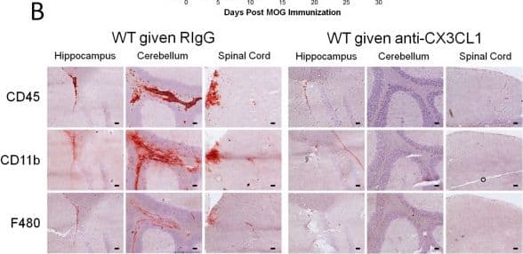
CX3CL1 antibody mediated blockade protects mice against EAE and its associated lymphocyte infiltration. Wild type mice were induced to develop EAE and starting at day 8 post induction given daily anti-CX3CL1 antibody or an isotype control treatments (i.p.). (A) EAE disease profile. Error bars represent the s.e.m. (n = 4 mice/group). Significant differences are indicated as determined by two-way ANOVA. EAE scoring data is representative of 2 separate experiments. (B) CD45, CD11b, and F480 stained brain (hippocampal and cerebellum areas) and spinal cord sections from day 28 post-EAE induced mice treated with either anti-CX3CL1 or control antibody. Positively stained cells (red) are shown against a hematoxylin counterstain (blue). Black scale bars represent 50 μm. (C) CD4 and (D) CD8 positive mean cells counts per field at 10x magnification from brain and spinal cord stained frozen brain sections from day 28 post-EAE induced mice treated with either anti-CX3CL1 or control antibody. Error bars represent the standard error of the mean (n ≤ 11). Significant differences (P < 0.05, *) are indicated as determined by the Student’s t-test. Image collected and cropped by CiteAb from the following publication (https://jneuroinflammation.biomedcentral.com/articles/10.1186/1742-2094-9-193), licensed under a CC-BY license. Not internally tested by R&D Systems.Applications for Mouse CX3CL1/Fractalkine Chemokine Domain Antibody
Application
Recommended Usage
Western Blot
1 µg/mL
Sample: Recombinant Mouse CX3CL1/Fractalkine Full Length (Catalog # 472-FF)
under non-reducing conditions only
Sample: Recombinant Mouse CX3CL1/Fractalkine Full Length (Catalog # 472-FF)
under non-reducing conditions only
Neutralization
Measured by its ability to neutralize CX3CL1/Fractalkine-induced chemotaxis in the BaF3 mouse pro-B cell line transfected with human CX3CR1. The Neutralization Dose (ND50) is typically 0.75-3.5 µg/mL in the presence of 30 ng/mL Recombinant Mouse CX3CL1/Fractalkine aa 25-105.
Mouse CX3CL1/Fractalkine Sandwich Immunoassay
ELISA Capture (Matched Antibody Pair)
Please Note: Optimal dilutions of this antibody should be experimentally determined.
Reviewed Applications
Read 1 review rated 4 using MAB571 in the following applications:
Formulation, Preparation, and Storage
Purification
Protein A or G purified from hybridoma culture supernatant
Reconstitution
Reconstitute at 0.5 mg/mL in sterile PBS. For liquid material, refer to CoA for concentration.
Formulation
Lyophilized from a 0.2 μm filtered solution in PBS with Trehalose. *Small pack size (SP) is supplied either lyophilized or as a 0.2 µm filtered solution in PBS.
Shipping
Lyophilized product is shipped at ambient temperature. Liquid small pack size (-SP) is shipped with polar packs. Upon receipt, store immediately at the temperature recommended below.
Stability & Storage
Use a manual defrost freezer and avoid repeated freeze-thaw cycles.
- 12 months from date of receipt, -20 to -70 °C as supplied.
- 1 month, 2 to 8 °C under sterile conditions after reconstitution.
- 6 months, -20 to -70 °C under sterile conditions after reconstitution.
Background: CX3CL1/Fractalkine
CX3CL1, also known as Fractalkine, is a type I membrane protein in which a chemokine domain possessing a unique C-X3-C cysteine motif is tethered on a long mucin-like stalk. It can also be released as a soluble molecule upon proteolysis at a membrane proximal site.
Alternate Names
FKN, Fractalkine, Neurotactin
Gene Symbol
CX3CL1
Additional CX3CL1/Fractalkine Products
Product Documents for Mouse CX3CL1/Fractalkine Chemokine Domain Antibody
Product Specific Notices for Mouse CX3CL1/Fractalkine Chemokine Domain Antibody
For research use only
Loading...
Loading...
Loading...
Loading...


