Mouse DPPIV/CD26 Antibody
R&D Systems, part of Bio-Techne | Catalog # MAB9541


Key Product Details
Species Reactivity
Validated:
Cited:
Applications
Validated:
Cited:
Label
Antibody Source
Product Specifications
Immunogen
Extracellular domain
Specificity
Clonality
Host
Isotype
Scientific Data Images for Mouse DPPIV/CD26 Antibody
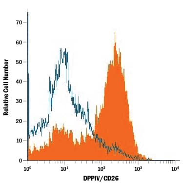
Detection of DPPIV/CD26 in Mouse Splenocytes by Flow Cytometry.
Mouse splenocytes were stained with Rat Anti-Mouse DPPIV/CD26 Monoclonal Antibody (Catalog # MAB9541, filled histogram) or isotype control antibody (Catalog # MAB006, open histogram), followed by Phycoerythrin-conjugated Anti-Rat IgG Secondary Antibody (Catalog # F0105B).Applications for Mouse DPPIV/CD26 Antibody
CyTOF-ready
Flow Cytometry
Sample: Mouse splenocytes
Formulation, Preparation, and Storage
Purification
Reconstitution
Formulation
Shipping
Stability & Storage
- 12 months from date of receipt, -20 to -70 °C as supplied.
- 1 month, 2 to 8 °C under sterile conditions after reconstitution.
- 6 months, -20 to -70 °C under sterile conditions after reconstitution.
Background: DPPIV/CD26
DPPIV/CD26 (EC 3.4.14.5) is a serine exopeptidase that releases Xaa-Pro dipeptides from the N-terminus of oligo- and polypeptides (1, 2). It is a type II membrane protein consisting of a short cytoplasmic tail, a transmembrane domain, and a long extracellular domain (3‑5). The extracellular domain contains glycosylation sites, a cysteine-rich region and the catalytic active site (Ser, Asp and His charge relay system). The amino acid sequence of the mouse DPPIV/CD26 extracellular domain is 84% and 91% identical to the human and rat counterparts, respectively. In the native state, DPPIV/CD26 is present as a noncovalently linked homodimer on the cell surface of a variety of cell types. The soluble form is also detectable in human serum and other body fluids, the levels of which may have clinical significance in patients with cancer, liver and kidney diseases, and depression. DPPIV/CD26 plays an important role in many biological and pathological processes. It functions as T cell-activating molecule (THAM). It serves as a co‑factor for entry of HIV in CD4+ cells (6). It binds adenosine deaminase, the deficiency of which causes severe combined immunodeficiency disease in humans (7). It cleaves chemokines such as stromal-cell-derived factor 1 alpha and macrophage-derived chemokine (8, 9). It degrades peptide hormones such as glucagon (10). It truncates procalcitonin, a marker for systemic bacterial infections with elevated levels detected in patients with thermal injury, sepsis and severe infection, and in children with bacterial meningitis (11).
References
- Misumi, Y. and Y. Ikehara (2004) in Handbook of Proteolytic Enzymes. Barrett, A.J. et al. (eds), p. 1905, Elsevier, London.
- Ikehara, Y. et al. (1994) Methods Enzymol. 244:215.
- Marguet, D. et al. (1992) J. Biol. Chem. 267:2200.
- Bernard, A.M. et al. (1994) Biochemistry 33:15204.
- Vivier, I. et al. (1991) J. Immunol. 147:447.
- Callebaut, C. et al. (1993) Science 262:2045.
- Kameoka, J. et al. (1993) Science 261:466.
- Ohtsuki, T. et al. (1998) FEBS Lett. 431:236.
- Proost, P. et al. (1999) J. Biol. Chem. 274:3988.
- Hinke, S.A. et al. (2000) J. Biol. Chem. 275:3827.
- Wrenger, S. et al. (2000) FEBS Lett. 466:155.
Long Name
Alternate Names
Gene Symbol
Additional DPPIV/CD26 Products
Product Documents for Mouse DPPIV/CD26 Antibody
Product Specific Notices for Mouse DPPIV/CD26 Antibody
For research use only