Mouse IL-1 beta/IL-1F2 Antibody
R&D Systems, part of Bio-Techne | Catalog # AB-401-NA

Key Product Details
Validated by
Biological Validation
Species Reactivity
Validated:
Mouse
Cited:
Mouse, Rat
Applications
Validated:
Neutralization, Simple Western, Western Blot
Cited:
Immunohistochemistry, Neutralization, Western Blot
Label
Unconjugated
Antibody Source
Polyclonal Goat IgG
Product Specifications
Immunogen
E. coli-derived recombinant mouse IL-1 beta/IL-1F2
Val118-Ser269
Accession # P10749
Val118-Ser269
Accession # P10749
Specificity
Detects mouse IL-1 beta/IL-1F2 in direct ELISAs and Western blots. In direct ELISAs, approximately 40% cross-reactivity with recombinant cotton rat IL-1 beta and recombinant rhesus monkey IL-1 beta is observed, approximately 15% cross-reactivity with recombinant rat IL-1 beta and recombinant rabbit IL-1 beta is observed, and less than 10% cross-reactivity with recombinant canine IL-1 beta, recombinant equine IL-1 beta, recombinant guinea pig IL-1 beta, recombinant porcine IL-1 beta, recombinant feline IL-1 beta, and recombinant human IL-1 beta is observed.
Clonality
Polyclonal
Host
Goat
Isotype
IgG
Endotoxin Level
<0.10 EU per 1 μg of the antibody by the LAL method.
Scientific Data Images for Mouse IL-1 beta/IL-1F2 Antibody
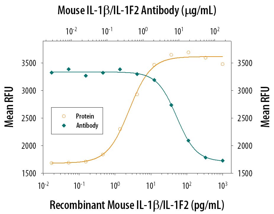
Cell Proliferation Induced by IL‑1 beta/IL‑1F2 and Neutral-ization by Mouse IL‑1 beta/IL‑1F2 Antibody.
Recombinant Mouse IL-1 beta/IL-1F2 (401-ML) stimulates proliferation in the the D10.G4.1 mouse helper T cell line in a dose-dependent manner (orange line). Proliferation elicited by Recombinant Mouse IL-1 beta/IL-1F2 (10 pg/mL) is neutralized (green line) by increasing concentrations of Goat Anti-Mouse IL-1 beta/IL-1F2 Polyclonal Antibody (Catalog # AB-401-NA). The ND50 is typically 2-12 µg/mL.Detection of Human and Mouse IL‑1 beta/IL‑1F2 by Western Blot.
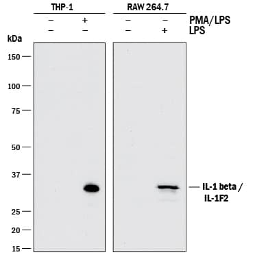
Western blot shows lysates of THP-1 human acute monocytic leukemia cell line untreated (-) or treated (+) with 200 nM PMA for 24 hours and 10 µg/mL LPS for 4 hours and RAW 264.7 mouse monocyte/macrophage cell line untreated (-) or treated (+) with 10 µg/mL LPS for 24 hours. PVDF membrane was probed with 1 µg/mL of Goat Anti-Mouse IL-1 beta/IL-1F2 Polyclonal Antibody (Catalog # AB-401-NA) followed by HRP-conjugated Anti-Goat IgG Secondary Antibody (HAF017). A specific band was detected for IL-1 beta/IL-1F2 at approximately 35 kDa (as indicated). This experiment was conducted under reducing conditions and using Immunoblot Buffer Group 1.Detection of Mouse IL‑1 beta/IL‑1F2 by Simple WesternTM.
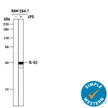
Simple Western lane view shows lysates of RAW 264.7 mouse monocyte/macrophage cell line untreated (-) or treated (+) with 10 µg/mL LPS for 24 hours, loaded at 0.5 mg/mL. A specific band was detected for IL‑1 beta/IL‑1F2 at approximately 40 kDa (as indicated) using 50 µg/mL of Goat Anti-Mouse IL‑1 beta/IL‑1F2 Polyclonal Antibody (Catalog # AB-401-NA) . This experiment was conducted under reducing conditions and using the 12-230 kDa separation system.Applications for Mouse IL-1 beta/IL-1F2 Antibody
Application
Recommended Usage
Simple Western
50 µg/mL
Sample: RAW 264.7 mouse monocyte/macrophage cell line treated with LPS
Sample: RAW 264.7 mouse monocyte/macrophage cell line treated with LPS
Western Blot
1 µg/mL
Sample: THP‑1 human acute monocytic leukemia cell line treated with PMA and LPS and RAW 264.7 mouse monocyte/macrophage cell line treated with LPS
Sample: THP‑1 human acute monocytic leukemia cell line treated with PMA and LPS and RAW 264.7 mouse monocyte/macrophage cell line treated with LPS
Neutralization
Measured by its ability to neutralize IL‑1 beta/IL‑1F2-induced proliferation in the D10.G4.1 mouse helper T cell line [Symons, J.A. et al. (1987) in Lymphokines and Interferons, a Practical Approach. Clemens, M.J. et al. (eds): IRL Press. 272]. The Neutralization Dose (ND50) is typically 2-12 µg/mL in the presence of 10 pg/mL Recombinant Mouse IL‑1 beta/IL‑1F2.
Formulation, Preparation, and Storage
Purification
Protein A or G purified
Reconstitution
Reconstitute at 1 mg/mL in sterile PBS.
Formulation
Lyophilized from a 0.2 μm filtered solution in PBS with Trehalose.
Shipping
The product is shipped at ambient temperature. Upon receipt, store it immediately at the temperature recommended below.
Stability & Storage
Use a manual defrost freezer and avoid repeated freeze-thaw cycles.
- 12 months from date of receipt, -20 to -70 °C as supplied.
- 1 month, 2 to 8 °C under sterile conditions after reconstitution.
- 6 months, -20 to -70 °C under sterile conditions after reconstitution.
Background: IL-1 beta/IL-1F2
Long Name
Interleukin 1 beta
Alternate Names
IL-1b, IL-1F2, IL1 beta, IL1B
Entrez Gene IDs
Gene Symbol
IL1B
UniProt
Additional IL-1 beta/IL-1F2 Products
Product Documents for Mouse IL-1 beta/IL-1F2 Antibody
Product Specific Notices for Mouse IL-1 beta/IL-1F2 Antibody
For research use only
Loading...
Loading...
Loading...
Loading...


