Mouse Leptin R Antibody
R&D Systems, part of Bio-Techne | Catalog # AF497


Key Product Details
Species Reactivity
Validated:
Cited:
Applications
Validated:
Cited:
Label
Antibody Source
Product Specifications
Immunogen
Ala20-Gly839
Accession # Q3US58
Specificity
Clonality
Host
Isotype
Scientific Data Images for Mouse Leptin R Antibody
Detection of Leptin R in Mouse Brain
Leptin R was detected in immersion fixed frozen sections of mouse brain using Goat Anti-Mouse Leptin R Antigen Affinity-purified Polyclonal Antibody (Catalog # AF497) at 1 µg/ml for 1 hour at room temperature followed by incubation with the Anti-Goat IgG VisUCyte™ HRP Polymer Antibody (Catalog # VC004). Tissue was stained using DAB (brown) and counterstained with hematoxylin (blue). Specific staining was localized to the cell surface. View our protocol for Chromogenic IHC Staining of Frozen Tissue Sections.Leptin R in Rat Brain.
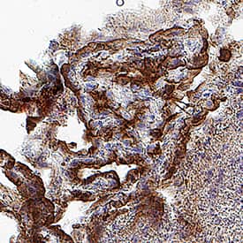
Leptin R was detected in perfusion fixed frozen sections of rat brain using 15 µg/mL Goat Anti-Mouse Leptin R Antigen Affinity-purified Polyclonal Antibody (Catalog # AF497) overnight at 4 °C. Tissue was stained with the Anti-Goat HRP-DAB Cell & Tissue Staining Kit (brown; Catalog # CTS008) and counterstained with hematoxylin (blue). Specific labeling was localized to the cytoplasm of cells in the choroid plexus. View our protocol for Chromogenic IHC Staining of Frozen Tissue Sections.Detection of Human Leptin R by Western Blot
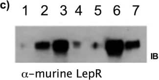
Expression and isolation of recombinant murine LepRec-Fc fusion proteins.a) Expression of murine LepRec-Fc constructs in adherent HEK293T/17 cells. Supernatants containing LepRec-Fc chimera proteins from 24 and 48 hours post-transfection were cleared of cellular debris, subjected to SDS PAGE and western blotted with antibodies against domains of murine IgG1 (Fc specific for lanes 1–5 and Ab specific for lanes 6–8). Lane 1: Mock transfected at 48 hours. Lane 2: WT transfected at 24 hours. Lane 3: WT transfected at 48 hours. Lane 4: Q223R transfected at 24 hours. Lane 5: Q223R transfected at 48 hours. Lane 6: Mock transfected at 48 hours. Lane 7: WT transfected at 48 hours. Lane 8: Q223R transfected at 48 hours. b) Concentration and buffer exchange of murine LepRec-Fc chimeras. Supernatants were collected 48 hours after growth medium was replaced with expression medium (serum free Optimem +2 mM sodium butyrate). These supernatant preparations were concentrated by Amicon ultrafiltration (NMWCO of 100 kDa). Supernatants, concentrates, and filtrates from each chimera were subjected to SDS-PAGE followed by coomassie staining (top) and western blotting with alpha-murine IgG1 specific to the Fc region (bottom). Lane 1: Expression medium. Lane 2: WT supernatant. Lane 3: WT concentrate. Lane 4: WT filtrate. Lane 5: Q223R supernatant. Lane 6: Q223R Concentrate. Lane 7: Q223R Filtrate. c) Supernatants, concentrates, and filtrates (as in b) from each chimera were subjected to SDS-PAGE followed by transfer to PVDF membrane and western blotting with alpha-murine leptin receptor (R&D scientific). Lane 1: Expression medium. Lane 2: WT supernatant. Lane 3: WT concentrate. Lane 4: WT filtrate. Lane 5: Q223R Filtrate. Lane 6: Q223R Concentrate. Lane 7: Q223R supernatant. Image collected and cropped by CiteAb from the following publication (https://pubmed.ncbi.nlm.nih.gov/24743494), licensed under a CC-BY license. Not internally tested by R&D Systems.Applications for Mouse Leptin R Antibody
CyTOF-ready
Flow Cytometry
Sample: Mouse splenocytes
Immunohistochemistry
Sample: Perfusion fixed frozen sections of rat and mouse brain
Western Blot
Sample: Recombinant Mouse Leptin R Fc Chimera (Catalog # 497-LR)
Reviewed Applications
Read 2 reviews rated 4.5 using AF497 in the following applications:
Formulation, Preparation, and Storage
Purification
Reconstitution
Formulation
*Small pack size (-SP) is supplied either lyophilized or as a 0.2 µm filtered solution in PBS.
Shipping
Stability & Storage
- 12 months from date of receipt, -20 to -70 °C as supplied.
- 1 month, 2 to 8 °C under sterile conditions after reconstitution.
- 6 months, -20 to -70 °C under sterile conditions after reconstitution.
Background: Leptin R
Leptin receptor (OB-R), also named B219, is a type I cytokine receptor family protein with significant amino acid sequence identity with gp130, G-CSF receptor, and the LIF receptor. Multiple isoforms of human and mouse OB-R, including a long form (OB-RL) with a large cytoplasmic domain capable of signal-transduction, and several receptor isoforms with short cytoplasmic domains (OB-Rs) lacking signal-transducing capabilities, have been identified. The extracellular domains of the short and long forms of OB-R are identical. An OB-R transcript lacking a transmembrane domain and potentially encoding a soluble form of the receptor has also been described. Circulating soluble OB-R, complexed to leptin, has been detected in mouse serum. Serum soluble OB-R levels have been shown to increase during pregnancy. OB-RL transcripts were reported to be expressed predominantly in regions of the hypothalamus previously thought to be important in body weight regulation. Expression of OB-Rs transcripts have been found in multiple tissues, including the choroid plexus, lung, kidney and primitive hematopoietic cell populations. OB-R has recently been shown to be encoded by the mouse diabetes (db) and rat fatty (fa) genes. Rodents homozygous for the db or fa mutations have been known to exhibit an obesity phenotype.
Mouse OB-R long form encodes a 1162 amino acid (aa) residue precursor protein with a 22 aa residue signal peptide, an 817 aa residue extracellular domain, a 21 aa residue transmembrane domain, and a 302 aa residue cytoplasmic domain. The extracellular domain of OB-R contains two hemopoietin receptor domains, a fibronectin type III domain and the WSXWS domain. Recombinant murine soluble OB-R has been shown to bind leptin with high affinity and is a potent leptin antagonist.
References
- Tartaglia, L.A. et al. (1995) Cell 83:1263.
- Cioffi, J.A. et al. (1996) Nature Medicine 2:585.
- Lee, J.I. and J.M. Friedman (1996) Nature 379:632.
- Tartaglia, L.A. (1997) J. Biol. Chem. 272:6093.
- Gavrilova, O. et al. (1997) J. Biol. Chem. 272:30546.
Long Name
Alternate Names
Gene Symbol
UniProt
Additional Leptin R Products
Product Documents for Mouse Leptin R Antibody
Product Specific Notices for Mouse Leptin R Antibody
For research use only

