Mouse Ly-6G/Ly-6C (Gr-1) Antibody
R&D Systems, part of Bio-Techne | Catalog # MAB1037


Conjugate
Catalog #
Key Product Details
Species Reactivity
Validated:
Mouse
Cited:
Human, Mouse, Bovine, Xenograft
Applications
Validated:
CyTOF-ready, Flow Cytometry, Immunocytochemistry, Immunohistochemistry, Immunoprecipitation
Cited:
Flow Cytometry, Immunocytochemistry, Immunodepletion, Immunohistochemistry, Immunohistochemistry-Frozen, Immunohistochemistry-Paraffin, Neutralization, Western Blot
Label
Unconjugated
Antibody Source
Monoclonal Rat IgG2B Clone # RB6-8C5
Product Specifications
Immunogen
The immunogen for this antibody is Ly-6G (Gr-1, Gr1).
Specificity
Detects Ly-6G/Ly-6C (Gr-1). Weak cross-reactivity with Ly-6C is observed.
Clonality
Monoclonal
Host
Rat
Isotype
IgG2B
Scientific Data Images for Mouse Ly-6G/Ly-6C (Gr-1) Antibody
Ly-6G/Ly-6C (Gr-1) in Mouse Spleno-cytes.
Ly-6G/Ly-6C (Gr-1) was detected in immersion fixed mouse splenocytes using Mouse Ly-6G/Ly-6C (Gr-1) Monoclonal Anti-body (Catalog # MAB1037) at 10 µg/mL for 3 hours at room temperature. Cells were stained using the NorthernLights™ 557-conjugated Anti-Rat IgG Secondary Antibody (red; Catalog # NL013) and counter-stained with DAPI (blue). View our protocol for Fluorescent ICC Staining of Non-adherent Cells.Ly-6G/Ly-6C (Gr-1) in Mouse Spleen.
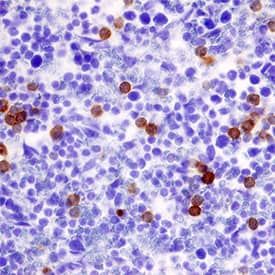
Ly-6G/Ly-6C (Gr-1) was detected in perfusion fixed frozen sections of mouse spleen using Rat Anti-Mouse Ly-6G/Ly-6C (Gr-1)Monoclonal Antibody (Catalog # MAB1037) at 3 µg/mL overnight at 4 °C. Tissue was stained using the Anti-Rat HRP-DAB Cell & Tissue Staining Kit (brown; Catalog # CTS017) and counterstained with hematoxylin (blue). Specific staining was localized to cytoplasm in splenocytes. View our protocol for Chromogenic IHC Staining of Frozen Tissue Sections.Detection of Mouse Mouse Ly-6G/Ly-6C (Gr-1) Antibody by Immunohistochemistry
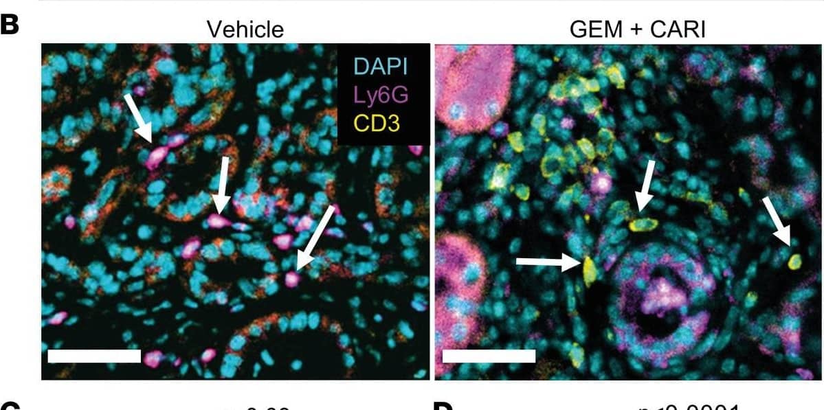
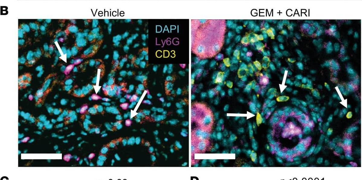
Lymphocyte/neutrophil ratio increases upon NHE1 inhibition in tumor sections of KPfC mice.(A) H&E (left) and PAS-stained KPfC mouse tissue sections after vehicle and gemcitabine + cariporide (GEM+CARI) therapy. Cells of innate immunity, such as neutrophils (arrows), utilize glycogen and are thus PAS+ (purple), in contrast to, for example, lymphocytes. Scale bar: 50 μm. (B) Representative IHC images stained for Ly6G+ neutrophils (magenta, arrows on left image), CD3+ lymphocytes (yellow, arrows on the right image), and nuclei with DAPI (cyan). Scale bar: 50 μm. (C) CD3/Ly6G ratio was assessed by dividing the number of CD3+ cells by the number of Ly6G+ cells in every tumor node. Data points depict the mean CD3/Ly6G ratio derived from each tumor node in individual mice; NVehicle = 10, NGEM = 9, NCARI = 10, NGEM+CARI = 11 mice. (D) To obtain the CD3/Ly6G ratio per tumor node, the number of CD3+ cells was divided by the respective number of Ly6G+ cells in each tumor node. Data points depict individual tumor nodes; nVehicle = 386, nGEM = 276, nCARI = 301, nGEM+CARI = 398. Data and statistical comparison in D and E are represented as median ± 95% CI using Kruskal-Wallis statistical test with Dunn’s post hoc test. Image collected and cropped by CiteAb from the following publication (https://pubmed.ncbi.nlm.nih.gov/37643024), licensed under a CC-BY license. Not internally tested by R&D Systems.Applications for Mouse Ly-6G/Ly-6C (Gr-1) Antibody
Application
Recommended Usage
CyTOF-ready
Ready to be labeled using established conjugation methods. No BSA or other carrier proteins that could interfere with conjugation.
Flow Cytometry
0.25 µg/106 cells
Sample: Mouse splenocytes or peripheral blood cells
Sample: Mouse splenocytes or peripheral blood cells
Immunocytochemistry
8-25 µg/mL
Sample: Immersion fixed mouse splenocytes
Sample: Immersion fixed mouse splenocytes
Immunohistochemistry
3-25 µg/mL
Sample: Perfusion fixed frozen sections of mouse spleen
Sample: Perfusion fixed frozen sections of mouse spleen
Immunoprecipitation
Conlan, J.W. and R.J. North (1994) J. Exp. Med. 179:259.
Reviewed Applications
Read 8 reviews rated 4.6 using MAB1037 in the following applications:
Formulation, Preparation, and Storage
Purification
Protein A or G purified from hybridoma culture supernatant
Reconstitution
Reconstitute at 0.5 mg/mL in sterile PBS. For liquid material, refer to CoA for concentration.
Formulation
Lyophilized from a 0.2 μm filtered solution in PBS with Trehalose. *Small pack size (SP) is supplied either lyophilized or as a 0.2 µm filtered solution in PBS.
Shipping
Lyophilized product is shipped at ambient temperature. Liquid small pack size (-SP) is shipped with polar packs. Upon receipt, store immediately at the temperature recommended below.
Stability & Storage
Use a manual defrost freezer and avoid repeated freeze-thaw cycles.
- 12 months from date of receipt, -20 to -70 °C as supplied.
- 1 month, 2 to 8 °C under sterile conditions after reconstitution.
- 6 months, -20 to -70 °C under sterile conditions after reconstitution.
Background: Ly-6G/Ly-6C (Gr-1)
References
- Spangrude, G.J. et al. (1988) Science 241:58.
- Fleming, T.J. et al. (1993) J. Immunol. 151:2399.
- Lewinsohn, D.M. et al.(1987) J. Immunol. 147:22.
- Lagasse, E. and I.L. Weissman (1996) J. Immunol. Methods 197:139.
Long Name
A Myeloid Differentiation Antigen
Alternate Names
Ly-6C, Ly-6G, Ly6G
Entrez Gene IDs
546644 (Mouse)
Gene Symbol
LY6G
Additional Ly-6G/Ly-6C (Gr-1) Products
Product Documents for Mouse Ly-6G/Ly-6C (Gr-1) Antibody
Product Specific Notices for Mouse Ly-6G/Ly-6C (Gr-1) Antibody
For research use only
Loading...
Loading...
Loading...
Loading...
Loading...


