Mouse Mast Cell Protease-1/Mcpt1 Antibody
R&D Systems, part of Bio-Techne | Catalog # AF5146

Key Product Details
Species Reactivity
Validated:
Cited:
Applications
Validated:
Cited:
Label
Antibody Source
Product Specifications
Immunogen
Glu19-Leu246
Accession # P11034
Specificity
Clonality
Host
Isotype
Scientific Data Images for Mouse Mast Cell Protease-1/Mcpt1 Antibody
Detection of Mouse Mast Cell Protease‑1/Mcpt1 by Western Blot.
Western blot shows lysates of mouse small intestine tissue. PVDF membrane was probed with 1 µg/mL of Sheep Anti-Mouse Mast Cell Protease-1/Mcpt1 Antigen Affinity-purified Polyclonal Antibody (Catalog # AF5146) followed by HRP-conjugated Anti-Sheep IgG Secondary Antibody (Catalog # HAF016). A specific band was detected for Mast Cell Protease-1/Mcpt1 at approximately 30 kDa (as indicated). This experiment was conducted under reducing conditions and using Immunoblot Buffer Group 1.Detection of Mouse Mast Cell Protease‑1/Mcpt1 by Simple WesternTM.
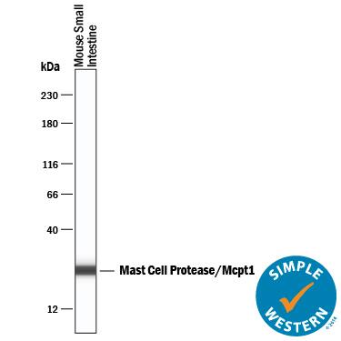
Simple Western lane view shows lysates of mouse small intestine tissue, loaded at 0.2 mg/mL. A specific band was detected for Mast Cell Protease-1/Mcpt1 at approximately 34 kDa (as indicated) using 50 µg/mL of Sheep Anti-Mouse Mast Cell Protease-1/Mcpt1 Antigen Affinity-purified Polyclonal Antibody (Catalog # AF5146) followed by 1:50 dilution of HRP-conjugated Anti-Sheep IgG Secondary Antibody (Catalog # HAF016). This experiment was conducted under reducing conditions and using the 12-230 kDa separation system.Applications for Mouse Mast Cell Protease-1/Mcpt1 Antibody
Simple Western
Sample: Mouse Small Intestine Tissue
Western Blot
Sample: Mouse Small Intestine Tissue
Formulation, Preparation, and Storage
Purification
Reconstitution
Formulation
Shipping
Stability & Storage
- 12 months from date of receipt, -20 to -70 °C as supplied.
- 1 month, 2 to 8 °C under sterile conditions after reconstitution.
- 6 months, -20 to -70 °C under sterile conditions after reconstitution.
Background: Mast Cell Protease-1/Mcpt1
Mast cell protease-1 (Mcpt1), also known as beta‑chymase, is a member of the Chymase family of chymotrypsin-like serine proteases (1). mMcpt1 is a mast cell protease predominantly expressed in intestinal mucosal mast cells where it promotes mucosal permeability in intestinal allergic hypersensitivity reactions (2). Its activation is completed by the removal of a two residue N‑terminal propeptide by a dipeptidyl peptidase (Cathepsin C) (3). Like human alpha‑Chymase, Mcpt1 is capable of the conversion of angiotensin I to angiotensin II, which plays a key role in the regulation of arterial pressure (4). Studies have shown that specific chymase inhibitors are able to diminish the development of abdominal aortic aneurysm and reduce the adhesion formation after cardiac surgery in hamsters (5, 6). Therefore, the development of specific inhibitors of chymase activity has been a pharmacologic strategy to develop therapeutic agents.
References
- Caughey, G.H. (2004) in Handbook of Proteolytic Enzymes (ed. Barrett, et al.), p. 1531 Academic Press, San Diego.
- Wastling, J.M. et al. (1998) Am. J. Pathol. 153:491.
- Murakami, M. et al. (1995) J. Biol. Chem. 270:2218.
- Saito, K. et al. (2003) Biochem. Biophys. Res. Commun. 302:773.
- Tsunemi, K. et al. (2004) J. Pharmacol. Exp. Ther. 309:879.
- Soga, Y. et al. (2004) J. Thorac. Cardiovasc. Surg. 127:72.
Alternate Names
Gene Symbol
UniProt
Additional Mast Cell Protease-1/Mcpt1 Products
Product Documents for Mouse Mast Cell Protease-1/Mcpt1 Antibody
Product Specific Notices for Mouse Mast Cell Protease-1/Mcpt1 Antibody
For research use only

