Mouse MSP/MST1 Antibody
R&D Systems, part of Bio-Techne | Catalog # AF6244

Key Product Details
Validated by
Species Reactivity
Applications
Label
Antibody Source
Product Specifications
Immunogen
Gly19-Gly716 (Cys677Ala)
Accession # NP_032269
Specificity
Clonality
Host
Isotype
Scientific Data Images for Mouse MSP/MST1 Antibody
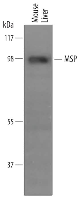
Detection of Mouse MSP/MST1 by Western Blot.
Western blot shows lysates of mouse liver tissue. PVDF membrane was probed with 1 µg/mL of Sheep Anti-Mouse MSP/MST1 Antigen Affinity-purified Polyclonal Antibody (Catalog # AF6244) followed by HRP-conjugated Anti-Sheep IgG Secondary Antibody (Catalog # HAF016). A specific band was detected for MSP/MST1 at approximately 96 kDa (as indicated). This experiment was conducted under reducing conditions and using Immunoblot Buffer Group 8.MSP/MST1 in Mouse Liver.
MSP/MST1 was detected in perfusion fixed frozen sections of mouse liver using Sheep Anti-Mouse MSP/MST1 Antigen Affinity-purified Polyclonal Antibody (Catalog # AF6244) at 15 µg/mL overnight at 4 °C. Tissue was stained using the Anti-Sheep HRP-DAB Cell & Tissue Staining Kit (brown; Catalog # CTS019) and counterstained with hematoxylin (blue). Specific staining was localized to hepatocytes. View our protocol for Chromogenic IHC Staining of Frozen Tissue Sections.Detection of Mouse MSP/MST1 by Western Blot
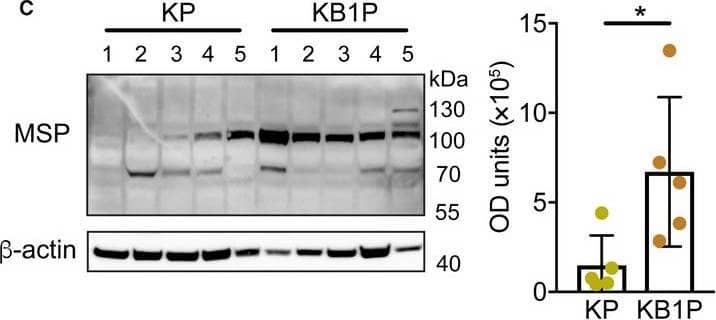
MSP stimulates AKT and MAPK signaling pathways through the RON receptor. (A) Mst1r mRNA expression assessed by qRT–PCR in cell lines derived from three KP tumors and four KB1P tumors, respectively. (B) Western blot analysis of RON protein expression in the same cell lines used in A. beta‐Actin was used as sample integrity control (same sample, different blot). (C) Western blot analysis of indicated proteins in the same KP and KB1P cell lines used in A, B. Cells were pretreated with 1 μm BMS‐777607 for 1 h prior to 100 ng·mL−1 recombinant MSP for 1 h where denoted. Images are representative of three replicate experiments. Total AKT and ERK were probed on the same blot as sample integrity controls. (D) Two KB1P cell lines were transduced with lentiviral shRNA vectors against Mst1r mRNA (shRON‐1 or shRON‐2) or control pLKO.1 vector. Confirmation of Mst1r mRNA knockdown quantified by qRT–PCR is expressed as relative to two housekeeping genes, Hprt and beta‐actin (each dot represents one technical replicate in a given experiment, the experiment was repeated three times for each cell line). (E) Two independent KB1P cell lines transduced with control or shRNA vectors against Mst1r mRNA were treated with 100 ng·mL−1 recombinant MSP for 1 h. Activation of AKT and ERK1/2 was assessed by western blot. Images are representative of three replicate transduction experiments. Data are represented as mean ± SD. **P < 0.01 as determined by one‐way ANOVA followed by Dunn’s post hoc test. n.s., not significant. Image collected and cropped by CiteAb from the following publication (https://pubmed.ncbi.nlm.nih.gov/32484599), licensed under a CC-BY license. Not internally tested by R&D Systems.Applications for Mouse MSP/MST1 Antibody
Immunohistochemistry
Sample: Perfusion fixed frozen sections of mouse liver
Western Blot
Sample: Mouse liver tissue
Formulation, Preparation, and Storage
Purification
Reconstitution
Formulation
Shipping
Stability & Storage
- 12 months from date of receipt, -20 to -70 °C as supplied.
- 1 month, 2 to 8 °C under sterile conditions after reconstitution.
- 6 months, -20 to -70 °C under sterile conditions after reconstitution.
Background: MSP/MST1
MSP (Macrophage stimulating protein 1; also hepatocyte growth factor-like protein) is an 80-95 kDa member of the peptidase S1 family of proteins. Although it is expressed principally by hepatocytes, it can also be induced in cells such as renal tubular epithelium. MSP has multiple targets, and as such, has multiple effects. It stimulates macrophage motility and phagocytosis, promotes keratinocyte and renal tubular cell proliferation, and depresses myeloid progenitor cell replication. Mouse proMSP is 698 amino acids (aa) in length. It contains one PAN (carbohydrate-binding) site (aa 19-105), four consecutive kringle domains (110-457) and an inactive peptidase S1 region (aa 489-714). An intrachain disulfide bond exists between Cys477 and Cys593 that becomes an interchain bond following protease cleavage between Arg488 and Val489. This creates a mature heterodimer containing a 45-57 kDa alpha-chain, and a 30-35 kDa beta-chain. Mouse proMSP shares 80% and 93% aa identity with human and rat proMSP, respectively.
Long Name
Alternate Names
Gene Symbol
UniProt
Additional MSP/MST1 Products
Product Documents for Mouse MSP/MST1 Antibody
Product Specific Notices for Mouse MSP/MST1 Antibody
For research use only


