Mouse Nanog Alexa Fluor® 488-conjugated Antibody
R&D Systems, part of Bio-Techne | Catalog # IC2729G


Key Product Details
Validated by
Species Reactivity
Applications
Label
Antibody Source
Product Specifications
Immunogen
Trp154-Leu262
Accession # Q80Z64
Specificity
Clonality
Host
Isotype
Scientific Data Images for Mouse Nanog Alexa Fluor® 488-conjugated Antibody
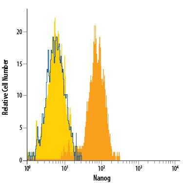
Detection of Nanog in D3 Mouse Cell Line by Flow Cytometry.
D3 mouse embryonic stem cell line either untreated (dark orange filled histogram) or treated with retinoic acid for 3 days (light orange filled histogram) was stained with Goat Anti-Mouse Nanog Alexa Fluor® 488-conjugated Antigen Affinity-purified Polyclonal Antibody (Catalog # IC2729G) or isotype control antibody (Catalog # IC108G, blue open histogram). To facilitate intracellular staining, cells were fixed with Flow Cytometry Fixation Buffer (Catalog # FC004) and permeabilized with Flow Cytometry Permeabilization/Wash Buffer I (Catalog # FC005). View our protocol for Staining Intracellular Molecules.Nanog in D3 Mouse Embryonic Stem Cells.
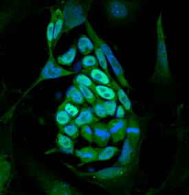
Nanog was detected in immersion fixed D3 mouse embryonic stem cell line on irradiated mouse embryonic fibroblasts using Goat Anti-Mouse Nanog Alexa Fluor® 488‑conjugated Antigen Affinity-purified Polyclonal Antibody (green; Catalog # IC2729G) at 10 µg/mL for 3 hours at room temperature. Cells were counterstained with DAPI (blue). Specific staining was localized to nuclei. View our protocol for Fluorescent ICC Staining of Stem Cells on Coverslips.Applications for Mouse Nanog Alexa Fluor® 488-conjugated Antibody
Immunocytochemistry
Sample: Immersion fixed D3 mouse embryonic stem cells on irradiated mouse embryonic fibroblasts
Intracellular Staining by Flow Cytometry
Sample: D3 mouse embryonic stem cell line fixed with Flow Cytometry Fixation Buffer (Catalog # FC004) and permeabilized with Flow Cytometry Permeabilization/Wash Buffer I (Catalog # FC005)
Formulation, Preparation, and Storage
Purification
Formulation
Shipping
Stability & Storage
- 12 months from date of receipt, 2 to 8 °C as supplied.
Background: Nanog
References
- Mitsui, K. et al. (2003) Cell 11:631.
- Chambers, I. et al. (2003) Cell 113:643.
- Hart, A.H. et al. (2004) Dev. Dyn. 230:187.
Long Name
Alternate Names
Gene Symbol
UniProt
Additional Nanog Products
Product Specific Notices for Mouse Nanog Alexa Fluor® 488-conjugated Antibody
This product is provided under an agreement between Life Technologies Corporation and R&D Systems, Inc, and the manufacture, use, sale or import of this product is subject to one or more US patents and corresponding non-US equivalents, owned by Life Technologies Corporation and its affiliates. The purchase of this product conveys to the buyer the non-transferable right to use the purchased amount of the product and components of the product only in research conducted by the buyer (whether the buyer is an academic or for-profit entity). The sale of this product is expressly conditioned on the buyer not using the product or its components (1) in manufacturing; (2) to provide a service, information, or data to an unaffiliated third party for payment; (3) for therapeutic, diagnostic or prophylactic purposes; (4) to resell, sell, or otherwise transfer this product or its components to any third party, or for any other commercial purpose. Life Technologies Corporation will not assert a claim against the buyer of the infringement of the above patents based on the manufacture, use or sale of a commercial product developed in research by the buyer in which this product or its components was employed, provided that neither this product nor any of its components was used in the manufacture of such product. For information on purchasing a license to this product for purposes other than research, contact Life Technologies Corporation, Cell Analysis Business Unit, Business Development, 29851 Willow Creek Road, Eugene, OR 97402, Tel: (541) 465-8300. Fax: (541) 335-0354.
For research use only
