Mouse Nanog Antibody
R&D Systems, part of Bio-Techne | Catalog # AF2729


Conjugate
Catalog #
Key Product Details
Species Reactivity
Validated:
Mouse
Cited:
Human, Mouse
Applications
Validated:
CyTOF-ready, Immunocytochemistry, Intracellular Staining by Flow Cytometry, Western Blot
Cited:
Chromatin Immunoprecipitation (ChIP), Flow Cytometry, Immunocytochemistry, Immunohistochemistry, Immunohistochemistry-Frozen, Immunohistochemistry-Paraffin, Western Blot
Label
Unconjugated
Antibody Source
Polyclonal Goat IgG
Product Specifications
Immunogen
E. coli-derived recombinant mouse Nanog
Trp154-Leu262
Accession # Q80Z64
Trp154-Leu262
Accession # Q80Z64
Specificity
Detects recombinant mouse Nanog in Western blots. In this format, approximately 50% cross-reactivity with recombinant human Nanog is observed.
Clonality
Polyclonal
Host
Goat
Isotype
IgG
Scientific Data Images for Mouse Nanog Antibody
Detection of Nanog in D3 Mouse Cell Line by Flow Cytometry.
D3 mouse embryonic stem cell line was stained with Goat Anti-Mouse Nanog Antigen Affinity-purified Polyclonal Antibody (Catalog # AF2729, filled histogram) or isotype control antibody (Catalog # AB-108-C, open histogram), followed by Allophycocyanin-conjugated Anti-Goat IgG Secondary Antibody (Catalog # F0108). To facilitate intracellular staining, cells were fixed with Flow Cytometry Fixation Buffer (Catalog # FC004) and permeabilized with Flow Cytometry Permeabilization/Wash Buffer I (Catalog # FC005).Nanog in D3 Mouse Cell Line.
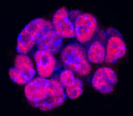
Nanog was detected in immersion fixed D3 mouse embryonic stem cell line using Goat Anti-Mouse Nanog Antigen Affinity-purified Polyclonal Antibody (Catalog # AF2729) at 10 µg/mL for 3 hours at room temperature. Cells were stained using the NorthernLights™ 557-conjugated Anti-Goat IgG Secondary Antibody (red; Catalog # NL001) and counterstained with DAPI (blue). Specific staining was localized to nuclei. View our protocol for Fluorescent ICC Staining of Stem Cells on Coverslips.Detection of Mouse Nanog by Immunocytochemistry/Immunofluorescence
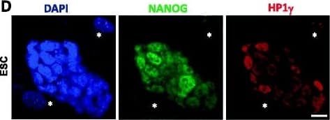
HP1 beta is highly expressed and diffuse in nuclei of pluripotent cells. a Confocal images of MEFs (top), R1 ESCs (middle) and Rr5 iPSCs (bottom) immunostained for Nanog (green, middle), HP1 beta (red, right) and counterstained with DAPI (blue, left). Asterisks indicate examples of MEFs used as a feeder layer in the culture of the pluripotent cells. b Quantification of the fluorescence intensities of Nanog (green bars) and HP1 beta (red bars) for the three cell types (n ≥ 26). Nanog is used as a marker for pluripotent cells; the fluorescence intensity of the background intensity was subtracted. c Number of HP1 beta foci in the different cell types. Error bars in (b) and (c) represent standard error of the mean. d Confocal images of R1 ESCs immunostained for Nanog (green, middle), HP1 gamma (red, right) and counterstained with DAPI (blue, left). e Confocal images of Rr5 iPSCs immunostained for HP1 gamma (red) and Nanog (inset, green). Asterisks indicate feeder layer MEF cells in (d) and (e). Scale bars for (a–e) = 15 μm. f Time lapse spinning disk confocal images of ESCs expressing the endogenous HP1 beta fused to mCherry induced to differentiate with 1 μM of retinoic acid (RA) for 40 hours (see also Additional file 7 for a video) Image collected and cropped by CiteAb from the following publication (https://genomebiology.biomedcentral.com/articles/10.1186/s13059-015-0760-8), licensed under a CC-BY license. Not internally tested by R&D Systems.Applications for Mouse Nanog Antibody
Application
Recommended Usage
CyTOF-ready
Ready to be labeled using established conjugation methods. No BSA or other carrier proteins that could interfere with conjugation.
Immunocytochemistry
5-15 µg/mL
Sample: Immersion fixed D3 mouse embryonic stem cell line
Sample: Immersion fixed D3 mouse embryonic stem cell line
Intracellular Staining by Flow Cytometry
0.25 µg/106 cells
Sample: D3 mouse embryonic stem cell line fixed with Flow Cytometry Fixation Buffer (Catalog # FC004) and permeabilized with Flow Cytometry Permeabilization/Wash Buffer I (Catalog # FC005)
Sample: D3 mouse embryonic stem cell line fixed with Flow Cytometry Fixation Buffer (Catalog # FC004) and permeabilized with Flow Cytometry Permeabilization/Wash Buffer I (Catalog # FC005)
Western Blot
Yang, W. et al. (2014) Nat. Comm. 5:3818
Formulation, Preparation, and Storage
Purification
Antigen Affinity-purified
Reconstitution
Reconstitute at 0.2 mg/mL in sterile PBS. For liquid material, refer to CoA for concentration.
Formulation
Lyophilized from a 0.2 μm filtered solution in PBS with Trehalose. *Small pack size (SP) is supplied either lyophilized or as a 0.2 µm filtered solution in PBS.
Shipping
Lyophilized product is shipped at ambient temperature. Liquid small pack size (-SP) is shipped with polar packs. Upon receipt, store immediately at the temperature recommended below.
Stability & Storage
Use a manual defrost freezer and avoid repeated freeze-thaw cycles.
- 12 months from date of receipt, -20 to -70 °C as supplied.
- 1 month, 2 to 8 °C under sterile conditions after reconstitution.
- 6 months, -20 to -70 °C under sterile conditions after reconstitution.
Background: Nanog
(1-3).
References
- Mitsui, K. et al. (2003) Cell 11:631.
- Chambers, I. et al. (2003) Cell 113:643.
- Hart, A.H. et al. (2004) Dev. Dyn. 230:187.
Long Name
Nanog Homeobox
Alternate Names
FLJ12581, FLJ40451, hNanog, homeobox protein NANOG, Homeobox transcription factor Nanog, homeobox transcription factor Nanog-delta 48, Nanog homeobox
Gene Symbol
NANOG
UniProt
Additional Nanog Products
Product Documents for Mouse Nanog Antibody
Product Specific Notices for Mouse Nanog Antibody
For research use only
Loading...
Loading...
Loading...
Loading...
Loading...


