Mouse NKp46/NCR1 Biotinylated Antibody
R&D Systems, part of Bio-Techne | Catalog # BAF2225


Key Product Details
Species Reactivity
Validated:
Cited:
Applications
Validated:
Cited:
Label
Antibody Source
Product Specifications
Immunogen
Glu22-Asn255
Accession # Q8C567
Specificity
Clonality
Host
Isotype
Scientific Data Images for Mouse NKp46/NCR1 Biotinylated Antibody
Detection of Mouse NKp46/NCR1 by Immunocytochemistry/Immunofluorescence
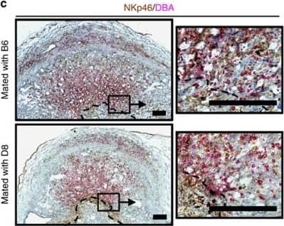
Paternal MHC expression on trophoblast.(a) Expression of paternal H-2Dd on gd9.5 trophoblast from B6 females impregnated by either B6 (top), D8 (middle) or BALB/c (bottom) males. BALB/c mice endogenously express H-2Dd. (b) H-2Dd and cytokeratin staining on control spleen cells. Scale bar=25 μm. (c) Representative sections of the distribution of NKp46+ DBA−(brown) and NKp46+ DBA+ (pink) uNK in the region of trophoblast invasion in the central decidua at gd9.5. Dashed lines demarcate the approximate boundary between invasive ectoplacental cone trophoblast and maternal decidua basalis. Scale bar=250 μm. DBA, Dolichos biflorus agglutinin. Image collected and cropped by CiteAb from the following publication (https://www.nature.com/articles/ncomms4359), licensed under a CC-BY license. Not internally tested by R&D Systems.Detection of Mouse NKp46/NCR1 by Immunocytochemistry/Immunofluorescence
Paternal H-2Dd expression on trophoblast results in impaired arterial remodelling.(a) Immunohistochemical staining for NKp46 and DBA reveals abundant uNK around decidual vessels, independent of paternal MHC. Scale bar=100 μm. DBA, Dolichos biflorus agglutinin. (b) H&E staining of uterine arteries illustrating the strategy used to measure vessel size (yellow dashed ring) and relative thickness (surface area ratio between areas surrounded by yellow and black ring, red arrows). Scale bar=25 μm. (c–e) Stereological and immunohistochemical analysis of arterial size (c) and relative thickness (d) in the decidua of B6 females mated with either B6 or D8 males. Pooled data from 4 litters per cross, n=13–15 implantation sites. P-values from unpaired Student’s t-tests. Means±s.e.m. (e) Representative staining for smooth muscle actin showing that the characteristic loss of actin in the media of spiral arteries is reduced in matings with D8 males (black arrows). Scale bars=100 μm. Image collected and cropped by CiteAb from the following publication (https://www.nature.com/articles/ncomms4359), licensed under a CC-BY license. Not internally tested by R&D Systems.Applications for Mouse NKp46/NCR1 Biotinylated Antibody
Western Blot
Sample: Recombinant Mouse NKp46/NCR1 Fc Chimera (Catalog # 2225-NK)
Reviewed Applications
Read 2 reviews rated 4.5 using BAF2225 in the following applications:
Formulation, Preparation, and Storage
Purification
Reconstitution
Formulation
Shipping
Stability & Storage
- 12 months from date of receipt, -20 to -70 °C as supplied.
- 1 month, 2 to 8 °C under sterile conditions after reconstitution.
- 6 months, -20 to -70 °C under sterile conditions after reconstitution.
Background: NKp46/NCR1
NKp46, along with NKp30 and NKp44, are activating receptors that have been collectively termed the natural cytotoxicity receptors (NCR) (1). These receptors are expressed almost exclusively by NK cells and play a major role in triggering some of the key lytic activities of NK cells. In human systems, the CD56dimCD16+ subpopulation that makes up the majority of NK cells in the peripheral blood and spleen expresses NKp46 in both resting and activated states (2). The main NK cell population of the lymph node (CD56brightCD16-) expresses low levels of NKp46 in resting cells, but expression is upregulated by IL-2. Mouse NKp46, also known as MAR-1 (3), is a type I transmembrane protein with two extracellular Ig-like domains. It has a positive charge in its transmembrane domain that permits association with the ITAM-bearing signal adapter proteins, CD3 zeta and Fc epsilon RI gamma (4). Studies with neutralizing antibodies indicate that the three NCR are primarily responsible for triggering the NK-mediated lysis of many human tumor cell lines. Blocking any of the NCRs individually resulted in partial inhibition of tumor cell lysis, but nearly complete inhibition of lysis was observed if all three receptors were blocked simultaneously (5). NKp46 has also been implicated in recognition of virus-infected cells through its capacity to bind to viral hemagglutinins (6 - 8).
References
- Moretta, L. and A. Moretta (2004) EMBO J. 23:255.
- Ferlazzo, G. et al. (2004) J. Immunol. 172:1455.
- Biassoni, R. et al. (1999) Eur. J. Immunol. 29:1014.
- Westgaard, I. et al. (2004) J. Leukoc. Biol. PMID 15356098.
- Pende, D. et al. (1999) J. Exp. Med. 190:1505.
- Arnon, T. et al. (2004) Blood 103:664.
- Arnon, T. et al. (2001) Eur. J. Immunol. 31:2680.
- Mandelboim, O. et al. (2001) Nature 409:1055.
Alternate Names
Gene Symbol
UniProt
Additional NKp46/NCR1 Products
Product Documents for Mouse NKp46/NCR1 Biotinylated Antibody
Product Specific Notices for Mouse NKp46/NCR1 Biotinylated Antibody
For research use only
