Mouse Noggin Antibody
R&D Systems, part of Bio-Techne | Catalog # AF719


Key Product Details
Species Reactivity
Validated:
Cited:
Applications
Validated:
Cited:
Label
Antibody Source
Product Specifications
Immunogen
Leu20-Cys232
Accession # P97466
Specificity
Clonality
Host
Isotype
Scientific Data Images for Mouse Noggin Antibody
Noggin in Embryonic Mouse Cardiac Tissue.
Noggin was detected in immersion fixed frozen sections of embryonic mouse cardiac tissue (11 d.p.c.) using 15 µg/mL Goat Anti-Mouse Noggin Antigen Affinity-purified Polyclonal Antibody (Catalog # AF719) overnight at 4 °C. Tissue was stained with the Anti-Goat HRP-DAB Cell & Tissue Staining Kit (brown; Catalog # CTS008) and counterstained with hematoxylin (blue). View our protocol for Chromogenic IHC Staining of Frozen Tissue Sections.Noggin in PC‑3 Human Cell Line.
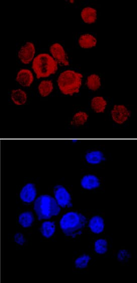
Noggin was detected in immersion fixed PC-3 human prostate cancer cell line using Goat Anti-Mouse Noggin Antigen Affinity-purified Polyclonal Antibody (Catalog # AF719) at 10 µg/mL for 3 hours at room temperature. Cells were stained using the NorthernLights™ 557-conjugated Anti-Goat IgG Secondary Antibody (red, upper panel; Catalog # NL001) and counterstained with DAPI (blue, lower panel). Specific staining was localized to cytoplasm. View our protocol for Fluorescent ICC Staining of Cells on Coverslips.Detection of Human Noggin by Immunohistochemistry
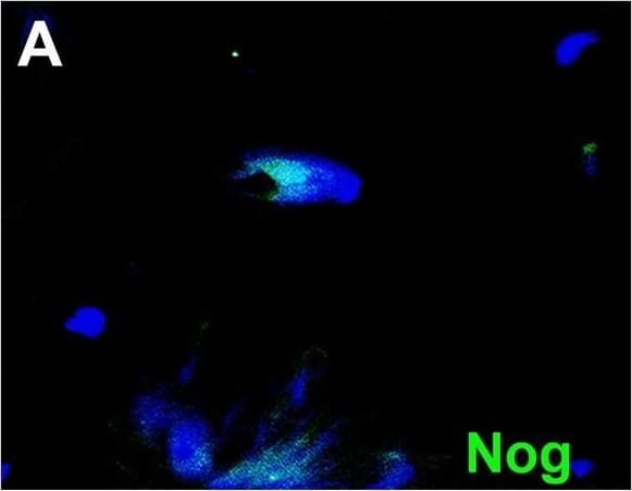
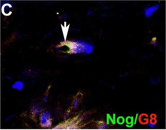
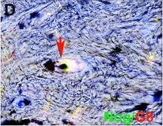
Myo/Nog cells contain ink in human tattooed skin.Tissue sections of tattooed skin were double labeled with the G8 mAb and antibodies to Noggin (Nog) (A-D), MyoD (F-I) or alpha-SMA (K-N). The colors of the secondary antibodies are indicated in the photographs. Nuclei were stained with Hoechst dye (blue). Images were produced with the epifluorescence (A-E, J and O) and confocal microscopes (F-I and K-N) with 60x lenses. Overlap of green and red fluorescence appears yellow in merged images (C, D, H, I, M and N). Fluorescent photomicrographs were merged with the corresponding DIC image to visualize the ink (black in D, I and N). Some double labeled cells appeared to contain ink (arrows in D, I and N). All ink laden G8+ cells were alpha-SMA+ (N). Smooth muscle cells of blood vessels also contained alpha-SMA (K). Minimal to no background fluorescence was visible after staining with the anti-goat (E), anti-IgM and anti-IgG (I), and anti-rabbit (O) secondary antibodies only. Bar = 28 μM in E and 5.6 μM in A-D and G-O. Image collected and cropped by CiteAb from the following open publication (https://pubmed.ncbi.nlm.nih.gov/32833999), licensed under a CC-BY license. Not internally tested by R&D Systems.Applications for Mouse Noggin Antibody
Immunocytochemistry
Sample: Immersion fixed PC‑3 human prostate cancer cell line
Immunohistochemistry
Sample: Immersion fixed frozen sections of embryonic mouse cardiac tissue (11 d.p.c.)
Western Blot
Sample: Recombinant Mouse Noggin (Catalog # 1967-NG)
Reviewed Applications
Read 5 reviews rated 4.8 using AF719 in the following applications:
Formulation, Preparation, and Storage
Purification
Reconstitution
Formulation
Shipping
Stability & Storage
- 12 months from date of receipt, -20 to -70 °C as supplied.
- 1 month, 2 to 8 °C under sterile conditions after reconstitution.
- 6 months, -20 to -70 °C under sterile conditions after reconstitution.
Background: Noggin
Noggin was originally cloned based on its dorsalizing activity in Xenopus embryos. Mammalian Noggins were subsequently identified and cloned from human, mouse and rat cDNA libraries. Mouse Noggin cDNA encodes a 232 amino acid (aa) residue precursor protein with 19 aa residue putative signal peptide that is cleaved to generate the 213 aa residue mature protein which is secreted as a homodimeric glycoprotein. Noggin is a highly conserved molecule. Mature mouse Noggin shares 99% and 83% aa sequence identity with human and Xenopus Noggin, respectively. Noggin has a complex pattern of expression during embryogenesis. In the adult, Noggin is expressed in the central nervous system and in several adult peripheral tissues such as lung, skeletal muscle and skin. Noggin has been shown to be a high-affinity BMP (bone morphogenetic protein) binding protein that antagonizes BMP bioactivities.
References
- Smith, W.C. and R.M. Harland (1992) Cell 70:829.
- Valenzuela, D.M. et al. (1995) J. Neurosci. 15:6077.
- Brunet, L.J. et al. (1998) Science 280:1455.
Alternate Names
Gene Symbol
UniProt
Additional Noggin Products
Product Documents for Mouse Noggin Antibody
Product Specific Notices for Mouse Noggin Antibody
For research use only


