Mouse PDGF-C Antibody
R&D Systems, part of Bio-Techne | Catalog # AF1447


Key Product Details
Species Reactivity
Validated:
Mouse
Cited:
Mouse, Rat
Applications
Validated:
Immunohistochemistry, Neutralization, Western Blot
Cited:
Immunohistochemistry, Immunohistochemistry-Paraffin, In vivo assay, Neutralization, Western Blot
Label
Unconjugated
Antibody Source
Polyclonal Goat IgG
Product Specifications
Immunogen
E. coli-derived recombinant mouse PDGF-CC
Val235-Gly345
Accession # Q8CI19
Val235-Gly345
Accession # Q8CI19
Specificity
Detects mouse PDGF‑C in direct ELISAs and Western blots. In direct ELISAs and Western blots, approximately 20% cross-reactivity with recombinant human (rh) PDGF-CC is observed and less than 1% cross-reactivity with rhPDGF-AA, rhPDGF-BB, rhPDGF-D, and recombinant rat PDGF-AB is observed.
Clonality
Polyclonal
Host
Goat
Isotype
IgG
Endotoxin Level
<0.10 EU per 1 μg of the antibody by the LAL method.
Scientific Data Images for Mouse PDGF-C Antibody
PDGF-C in Mouse Kidney.
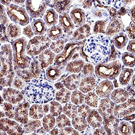
PDGF-C was detected in perfusion fixed paraffin-embedded sections of mouse kidney using Goat Anti-Mouse PDGF-C Antigen Affinity-purified Polyclonal Antibody (Catalog # AF1447) at 5 µg/mL for 1 hour at room temperature followed by incubation with the Anti-Goat IgG VisUCyte™ HRP Polymer Antibody (VC004). Before incubation with the primary antibody, tissue was subjected to heat-induced epitope retrieval using Antigen Retrieval Reagent-Basic (CTS013). Tissue was stained using DAB (brown) and counterstained with hematoxylin (blue). Specific staining was localized to cytoplasm and plasma membrane in convoluted tubules. Staining was performed using our protocol for IHC Staining with VisUCyte HRP Polymer Detection Reagents.Cell Proliferation Induced by PDGF‑CC and Neutralization by Mouse PDGF‑C Antibody.
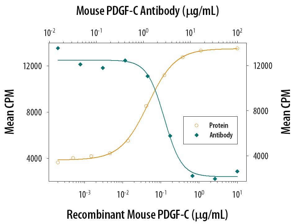
Recombinant Mouse PDGF‑CC stimulates proliferation in the NR6R‑3T3 mouse fibroblast cell line in a dose-dependent manner (orange line). Proliferation elicited by 0.8 µg/mL Recombinant Mouse PDGF‑CC is neutralized (green line) by increasing concentrations of Goat Anti-Mouse PDGF-C Antigen Affinity-purified Poly-clonal Antibody (Catalog # AF1447). The ND50 is typically 4‑16 µg/mL.Detection of Mouse PDGF-C by Immunocytochemistry/ Immunofluorescence
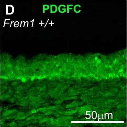
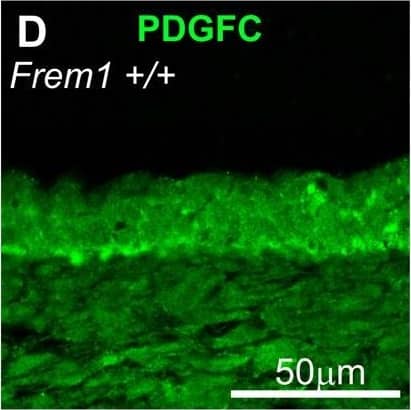
FREM1 regulates activation of AKT and MAPK upon PDGFC stimulation. (A) Representative western blotting of phosphorylation of AKT and MAPK ERK1/2 in MEFs from WT and bat mouse embryos stimulated with PDGFCC for the indicated time periods. (B) Relative quantification of AKT phosphorylation levels. WT cells 10 minutes after stimulation were assigned a value of 1 and all other samples are standardised against this value. Graph represents average of up to nine WT and 16 bat samples, performed across four independent experiments from at least three different cell lines for each genotype. Black bars: WT; white bars, bat mutant. (C) FREM1 mutation in bat mutants reduces phosphorylation of PDGFR alpha in response to the addition of exogenous PDGFCC. IP, immunoprecipitation antibody; WB, western blotting antibody. (D) E13.5 WT embryo head skin sections stained for PDGFC (green), PDGFR alpha (red) and nuclear dye DAPI (blue). Error bars represent standard error of the mean (s.e.m.); *P<0.05, **P<0.01, ***P<0.005. Image collected and cropped by CiteAb from the following open publication (https://pubmed.ncbi.nlm.nih.gov/24046351), licensed under a CC-BY license. Not internally tested by R&D Systems.Applications for Mouse PDGF-C Antibody
Application
Recommended Usage
Immunohistochemistry
5-15 µg/mL
Sample: Perfusion fixed paraffin-embedded sections of mouse kidney
Sample: Perfusion fixed paraffin-embedded sections of mouse kidney
Western Blot
0.1 µg/mL
Sample: Recombinant Mouse PDGF-C
Sample: Recombinant Mouse PDGF-C
Neutralization
Measured by its ability to neutralize PDGF-CC-induced proliferation in the NR6R-3T3 mouse fibroblast cell line. Raines, E. W. et al. (1985) Methods Enzymol. 109:749. The Neutralization Dose (ND50) is typically 4-16 µg/mL in the presence of 0.8 µg/mL Recombinant Mouse PDGF-CC.
Formulation, Preparation, and Storage
Purification
Antigen Affinity-purified
Reconstitution
Reconstitute at 0.2 mg/mL in sterile PBS. For liquid material, refer to CoA for concentration.
Formulation
Lyophilized from a 0.2 μm filtered solution in PBS with Trehalose. *Small pack size (SP) is supplied either lyophilized or as a 0.2 µm filtered solution in PBS.
Shipping
Lyophilized product is shipped at ambient temperature. Liquid small pack size (-SP) is shipped with polar packs. Upon receipt, store immediately at the temperature recommended below.
Stability & Storage
Use a manual defrost freezer and avoid repeated freeze-thaw cycles.
- 12 months from date of receipt, -20 to -70 °C as supplied.
- 1 month, 2 to 8 °C under sterile conditions after reconstitution.
- 6 months, -20 to -70 °C under sterile conditions after reconstitution.
Background: PDGF-C
Long Name
Platelet-derived Growth Factor C
Alternate Names
PDGFC
Gene Symbol
PDGFC
UniProt
Additional PDGF-C Products
Product Documents for Mouse PDGF-C Antibody
Product Specific Notices for Mouse PDGF-C Antibody
For research use only
Loading...
Loading...
Loading...
Loading...


