Mouse/Rat Inositol Monophosphatase 3/IMPAD1 Antibody
R&D Systems, part of Bio-Techne | Catalog # AF7028

Key Product Details
Species Reactivity
Validated:
Cited:
Applications
Validated:
Cited:
Label
Antibody Source
Product Specifications
Immunogen
Asn145-His356
Accession # Q80V26
Specificity
Clonality
Host
Isotype
Scientific Data Images for Mouse/Rat Inositol Monophosphatase 3/IMPAD1 Antibody
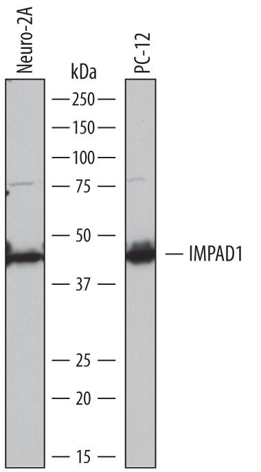
Detection of Mouse and Rat Inositol Monophosphatase 3/IMPAD1 by Western Blot.
Western blot shows lysates of Neuro-2A mouse neuroblastoma cell line and PC-12 rat adrenal pheochromocytoma cell line. PVDF membrane was probed with 1 µg/mL of Sheep Anti-Mouse Inositol Monophosphatase 3/IMPAD1 Antigen Affinity-purified Polyclonal Antibody (Catalog # AF7028) followed by HRP-conjugated Anti-Sheep IgG Secondary Antibody (Catalog # HAF016). A specific band was detected for Inositol Monophosphatase 3/IMPAD1 at approximately 42 kDa (as indicated). This experiment was conducted under reducing conditions and using Immunoblot Buffer Group 1.Inositol Monophosphatase 3/IMPAD1 in Mouse Mesenchymal Stem Cells.
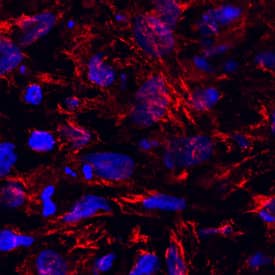
Inositol Monophosphatase 3/IMPAD1 was detected in immersion fixed mouse mesenchymal stem cells differentiated into chondrocytes using Sheep Anti-Mouse/Rat Inositol Monophosphatase 3/IMPAD1 Antigen Affinity-purified Polyclonal Antibody (Catalog # AF7028) at 10 µg/mL for 3 hours at room temperature. Cells were stained using the NorthernLights™ 557-conjugated Anti-Sheep IgG Secondary Antibody (red; Catalog # NL010) and counterstained with DAPI (blue). Specific staining was localized to cytoplasm. View our protocol for Fluorescent ICC Staining of Cells on Coverslips.Applications for Mouse/Rat Inositol Monophosphatase 3/IMPAD1 Antibody
Immunocytochemistry
Sample: Immersion fixed mouse mesenchymal stem cells differentiated into chondrocytes
Western Blot
Sample: Neuro‑2A mouse neuroblastoma cell line and PC‑12 rat adrenal pheochromocytoma cell line
Formulation, Preparation, and Storage
Purification
Reconstitution
Formulation
Shipping
Stability & Storage
- 12 months from date of receipt, -20 to -70 °C as supplied.
- 1 month, 2 to 8 °C under sterile conditions after reconstitution.
- 6 months, -20 to -70 °C under sterile conditions after reconstitution.
Background: Inositol Monophosphatase 3/IMPAD1
IMPAD1 (Inositol monophosphatase domain-containing protein 1; also IMPA3, gPAPP and IMPase 3) is a 40-42 kDa member of the inositol monophosphatase family of proteins. It is expressed in embryo, and found in Purkinje cells, brain stem, lung and chondrocytes. IMPAD1 in theory may catalyze the synthesis of myo-inositol from myo-inositol monophosphate. Free myo-inositol is used to generate inositol phospholipid, an essential component of intracellular signaling pathways that mobilize calcium. IMPAD1 is reported to promote sulfation of chondroitin by converting PAP (an endproduct of the sulfation process) to 5'-AMP. PAP is a known inhibitor of SULTs. IMPAD1 is a 356 amino acid (aa) type II transmembrane Golgi-embedded glycoprotein. It contains a short cytoplasmic tail (aa 1-12) and an extended luminal region (aa 34-356) that contains its catalytic domain (aa 60-347). Over aa 145-356, mouse IMPAD1 shares 99% and 93% aa sequence identity with rat and human IMPAD1, respectively.
Alternate Names
Gene Symbol
UniProt
Additional Inositol Monophosphatase 3/IMPAD1 Products
Product Documents for Mouse/Rat Inositol Monophosphatase 3/IMPAD1 Antibody
Product Specific Notices for Mouse/Rat Inositol Monophosphatase 3/IMPAD1 Antibody
For research use only

