Mouse Renin 1 Antibody
R&D Systems, part of Bio-Techne | Catalog # AF4277


Key Product Details
Species Reactivity
Validated:
Cited:
Applications
Validated:
Cited:
Label
Antibody Source
Product Specifications
Immunogen
Leu22-Arg402
Accession # P06281
Specificity
Clonality
Host
Isotype
Scientific Data Images for Mouse Renin 1 Antibody
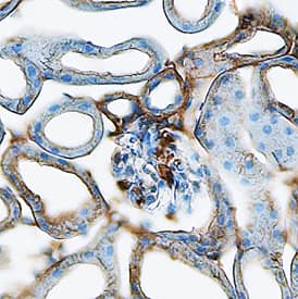
Renin in Mouse Kidney.
Renin was detected in perfusion fixed frozen sections of mouse kidney using Goat Anti-Mouse Renin 1 Antigen Affinity-purified Polyclonal Antibody (Catalog # AF4277) at 15 µg/mL overnight at 4 °C. Tissue was stained using the Anti-Goat HRP-DAB Cell & Tissue Staining Kit (brown; Catalog # CTS008) and counterstained with hematoxylin (blue). Specific staining was localized to cytoplasm in glomeruli and tubules. View our protocol for Chromogenic IHC Staining of Frozen Tissue Sections.Detection of Mouse Renin by Immunocytochemistry/Immunofluorescence
RLCs invade the glomerulus during EC model.A. Intraglomerular RLCs tagged by beta-gal, but negative for renin appear in the regenerative phase of the EC model (day 7). Representative confocal microscopy images for day 0 and day 7 of beta-gal/renin co-stained kidney slices. 4′,6-diamidino-2-phenylindole (DAPI) was used as a nuclear marker. The channels for green ( beta-gal) and red (renin) fluorescent signals in the dashed square on day 7 are separately shown in the small right panels. Scale bars correspond to 25 μm; B. Representative 3D reconstruction of glomeruli and beta-gal labelled RLCs (blue) on day 0 and day 7 of the EC model. The mesangial cell marker alpha8-integrin (red) was used to visualize the glomeruli. Scale bars correspond to 20 μm; C. Quantification of glomeruli with tufts containing beta-gal expressing RLCs in the regenerative phase of the EC model (day 7). Data are presented as mean ± SEM, n = 5/10 for day 0 (baseline) /day 7, respectively. n.d.—not detectable; D. The intraglomerular RLCs observed during the EC model are not of hematopoietic origin. Representative confocal microscopy images for day 0 and day 7 of beta-gal/CD45 (hematopoietic marker) co-stained kidney slices. 4′,6-diamidino-2-phenylindole (DAPI) was used as a nuclear marker. The channels for green ( beta-gal) and red (CD45) fluorescent signals in the dashed square on day 7 are separately shown in the small right panels. Scale bars correspond to 25 μm. Image collected and cropped by CiteAb from the following publication (https://pubmed.ncbi.nlm.nih.gov/29771991), licensed under a CC-BY license. Not internally tested by R&D Systems.Applications for Mouse Renin 1 Antibody
Immunohistochemistry
Sample: Perfusion fixed frozen sections of mouse kidney
Immunoprecipitation
Sample: Conditioned cell culture medium spiked with Recombinant Mouse Renin (Catalog # 4277-AS), see our available Western blot detection antibodies
Western Blot
Sample: Recombinant Mouse Renin (Catalog # 4277-AS)
Mouse Renin 1 Sandwich Immunoassay
Formulation, Preparation, and Storage
Purification
Reconstitution
Formulation
Shipping
Stability & Storage
- 12 months from date of receipt, -20 to -70 °C as supplied.
- 1 month, 2 to 8 °C under sterile conditions after reconstitution.
- 6 months, -20 to -70 °C under sterile conditions after reconstitution.
Background: Renin
Mouse Renin is a secreted, 42‑47 kDa glycosylated member of the peptidase A1 family. It is an aspartyl protease that cleaves angiotensinogen to form angiotensin I. In mouse, there are two genes that code for Renin. One is in the submandibular gland and the other is in the kidney. The two mature Renin molecules are 95% amino acid (aa) identical. Renal Renin (Renin 1) is synthesized as a 381 aa proform (aa 22‑402). In the kidney, pro‑Renin is proteolytically cleaved after Thr71 to generate a mature enzyme. Mouse pro Renin shares 70% and 85% aa sequence identity with human and rat pro Renin, respectively.
Alternate Names
Gene Symbol
UniProt
Additional Renin Products
Product Documents for Mouse Renin 1 Antibody
Product Specific Notices for Mouse Renin 1 Antibody
For research use only
