MyoD Antibody (5.8A) - BSA Free
Novus Biologicals, part of Bio-Techne | Catalog # NB100-56511

![Immunocytochemistry/ Immunofluorescence: MyoD Antibody (5.8A) - BSA Free [NB100-56511] Immunocytochemistry/ Immunofluorescence: MyoD Antibody (5.8A) - BSA Free [NB100-56511]](https://resources.bio-techne.com/images/products/MyoD-Antibody-5-8A-Immunocytochemistry-Immunofluorescence-NB100-56511-img0008.jpg)
Key Product Details
Validated by
Species Reactivity
Validated:
Cited:
Predicted:
Applications
Validated:
Cited:
Label
Antibody Source
Format
Concentration
Product Specifications
Immunogen
Specificity
Clonality
Host
Isotype
Description
Scientific Data Images for MyoD Antibody (5.8A) - BSA Free
Immunocytochemistry/ Immunofluorescence: MyoD Antibody (5.8A) - BSA Free [NB100-56511]
Immunocytochemistry/Immunofluorescence: MyoD Antibody (5.8A) [NB100-56511] - Human myoblasts were stained with MyoD1 antibody, diluted 1:200. ICC/IF image submitted by a verified customer review.Western Blot: MyoD Antibody (5.8A)BSA Free [NB100-56511]
Western Blot: MyoD Antibody (5.8A) [NB100-56511] - Insuline-like growth factor (IGF1) antagonized the effects of miR-106a-5p on myogenic differentiation in C2C12. All data were collected from C2C12 myotubes 5 d post differentiation. Western-blot analysis of myogenic regulatory factors (MyoD, MyoG, MyHC) in cells; (H) The statistical results of Figure 3G. Data were presented as mean +/- SEM. n = 3 per group. * p < 0.05, ** p < 0.01. Image collected and cropped by CiteAb from the following publication (https://www.mdpi.com/2073-4425/9/7/333/htm) licensed under a CC-BY license. anti-MyoG (catalog# NB100-56510)Immunocytochemistry/ Immunofluorescence: MyoD Antibody (5.8A) - BSA Free [NB100-56511]
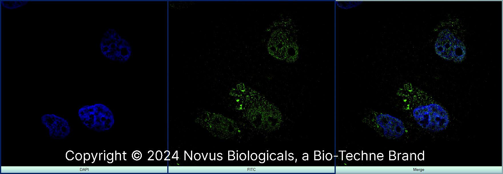
Immunocytochemistry/Immunofluorescence: MyoD Antibody (5.8A) [NB100-56511] - Cell Lines Tested: mouse skeletal muscle-derived primary cell population Test Sample Preparation: mouse skeletal muscle digested by collagenase type 2 System: Super Sensitive High Resolution Confocal Laser Microscope (LSM880 with Airyscan) Excitation Wavelength: 488nm Emission Filter: 562 nm. Image using the DyLight 488 form of this antibody. ICC/IF image submitted by a verified customer review.Applications for MyoD Antibody (5.8A) - BSA Free
Immunocytochemistry/ Immunofluorescence
Immunohistochemistry
Immunohistochemistry-Frozen
Immunoprecipitation
Western Blot
Reviewed Applications
Read 2 reviews rated 4 using NB100-56511 in the following applications:
Formulation, Preparation, and Storage
Purification
Formulation
Format
Preservative
Concentration
Shipping
Stability & Storage
Background: MyoD
Long Name
Alternate Names
Gene Symbol
UniProt
Additional MyoD Products
Product Documents for MyoD Antibody (5.8A) - BSA Free
Product Specific Notices for MyoD Antibody (5.8A) - BSA Free
There is considerable literature published using the MyoD, Clone 5.8A antibody. The original development publication of the MyoD antibody, Clone 5.8A showed that the antibody detected MyoD in rhabdomysosarcomas by IHC (frozen) but not in normal adult tissues (Dias, 1992) or normal fetal skeletal muscle. The 5.8A clone also detected MyoD1 in a subset of Wilms' tumors and one ectomesenchyoma, neoplasms known to contain myogenic elements. These results led to the concept in 1992 that the 5.8A clone may be useful for differentiating rhabdomyosarcomas from other soft tissue malignancies. However, as there has been a myriad of publications since Clone 5.8A was first described, users are encourage to consult the scientific literature citing Clone 5.8A to determine the suitability of the antibody for their model system.
This product is for research use only and is not approved for use in humans or in clinical diagnosis. Primary Antibodies are guaranteed for 1 year from date of receipt.
![Western Blot: MyoD Antibody (5.8A)BSA Free [NB100-56511] Western Blot: MyoD Antibody (5.8A)BSA Free [NB100-56511]](https://resources.bio-techne.com/images/products/MyoD-Antibody-5-8A-Western-Blot-NB100-56511-img0009.jpg)
![Immunocytochemistry/ Immunofluorescence: MyoD Antibody (5.8A) - BSA Free [NB100-56511] Immunocytochemistry/ Immunofluorescence: MyoD Antibody (5.8A) - BSA Free [NB100-56511]](https://resources.bio-techne.com/images/products/MyoD-Antibody-5-8A-Immunocytochemistry-Immunofluorescence-NB100-56511-img0007.jpg)
![Western Blot: MyoD Antibody (5.8A)BSA Free [NB100-56511] Western Blot: MyoD Antibody (5.8A)BSA Free [NB100-56511]](https://resources.bio-techne.com/images/products/MyoD-Antibody-5-8A-Western-Blot-NB100-56511-img0006.jpg)
![Western Blot: MyoD Antibody (5.8A)BSA Free [NB100-56511] Western Blot: MyoD Antibody (5.8A)BSA Free [NB100-56511]](https://resources.bio-techne.com/images/products/MyoD-Antibody-5-8A-Western-Blot-NB100-56511-img0010.jpg)
![Immunohistochemistry-Frozen: MyoD Antibody (5.8A) - BSA Free [NB100-56511] Immunohistochemistry-Frozen: MyoD Antibody (5.8A) - BSA Free [NB100-56511]](https://resources.bio-techne.com/images/products/MyoD-Antibody-5-8A-Immunohistochemistry-NB100-56511-img0005.jpg)
![Immunocytochemistry/ Immunofluorescence: MyoD Antibody (5.8A) - BSA Free [NB100-56511] Immunocytochemistry/ Immunofluorescence: MyoD Antibody (5.8A) - BSA Free [NB100-56511]](https://resources.bio-techne.com/images/products/MyoD-Antibody-5-8A-Immunocytochemistry-Immunofluorescence-NB100-56511-img0004.jpg)


