Rat CNTFR alpha Antibody
R&D Systems, part of Bio-Techne | Catalog # AF-559-NA


Key Product Details
Validated by
Species Reactivity
Validated:
Cited:
Applications
Validated:
Cited:
Label
Antibody Source
Product Specifications
Immunogen
Ala19-Pro346
Accession # Q08406
Specificity
Clonality
Host
Isotype
Endotoxin Level
Scientific Data Images for Rat CNTFR alpha Antibody
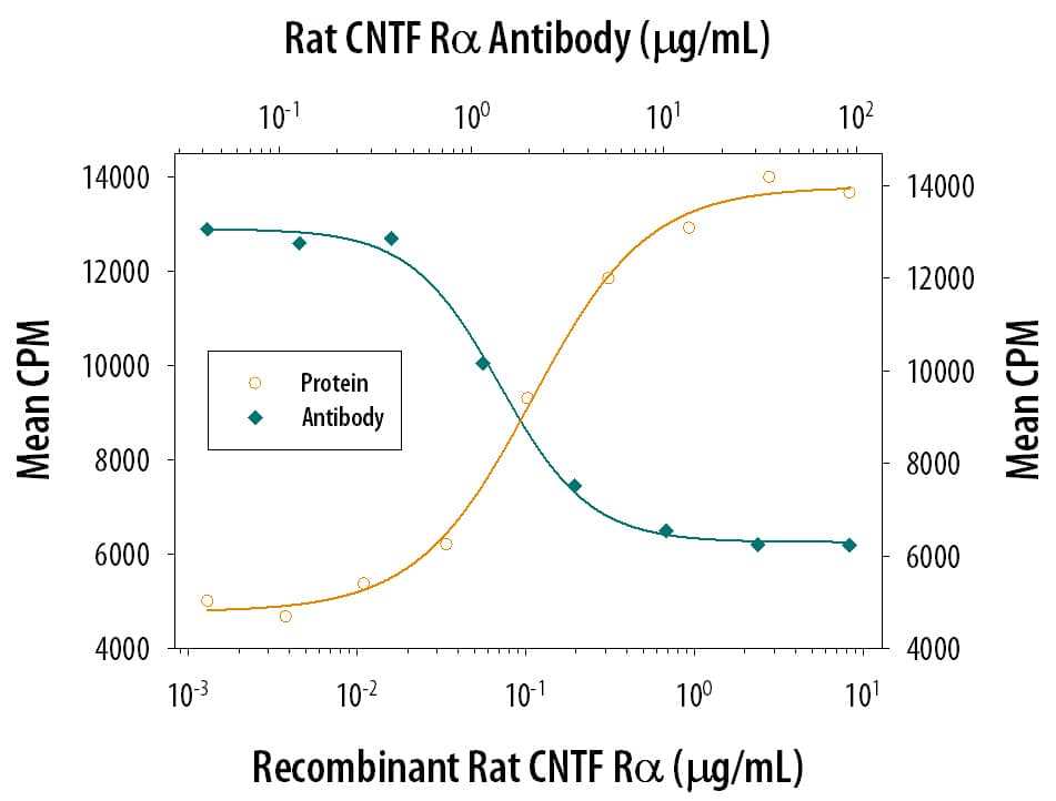
CNTF R alpha Enhancement of CNTF-dependent Cell Proliferation and Neutralization by Rat CNTF R alpha Antibody.
Recombinant Rat CNTF Ra (Catalog # 558-CR) enhances Recombinant Rat CNTF (Catalog # 557-NT) induced proliferation in the TF-1 human erythroleukemic cell line in a dose-dependent manner (orange line). Enhancement of Recombinant Rat CNTF (10 ng/mL) activity elicited by Recombinant Rat CNTF Ra (0.25 µg/mL) is neutralized (green line) by increasing concentrations of Goat Anti-Rat CNTF Ra Antigen Affinity-purified Polyclonal Antibody (Catalog # AF-559-NA). The ND50 is typically 2-6 µg/mL.Detection of Mouse CNTFR alpha by Immunocytochemistry/ Immunofluorescence
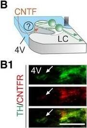
CNTF activates tyrosine hydroxylase by phosphorylation in mouse and human locus coeruleusCntfr mRNA expression in mouse locus coeruleus (LC, white arrows). Increasing mRNA content is indicated by a color gradient ranging from dark blue (low) to red (high). The ependymal surface as contact point with the 4th ventricle is shown in blue. Scale bar = 100 μm.Hypothetical orientation of NE neurons with their dendrites emanating toward the 4th ventricle. (B1) CNTF receptor (CNTFR) localization in tyrosine hydroxylase (TH)+ processes extending toward the ventricular wall (arrow marks ventricular surface). Scale bar = 10 μm.c‐Fos activation in LC 2 h after formalin stress. Left: Increased density of c‐Fos+ cells in the LC. Scale bar = 150 μm. Right: Quantitative data. *P < 0.05, n = 4/group.Representative Western blots of ex vivo LC explants acutely exposed to recombinant CNTF (for 20 min) show increased TH (D1) and Erk1 (D2) phosphorylation (n ≥ 3 samples/group). *P < 0.05.Intracerebroventricular infusion of Cntf siRNA blunts stress‐induced TH phosphorylation in the LC. (E1) Acute stress was triggered by injecting 4% PFA into a left hind paw unilaterally. Sampling followed 20 min later. *P < 0.05, n = 8/group.Schematic diagram of the BDA‐based retrograde labeling of LC neurons projecting to the mPFC. (F1) Multiple‐labeling immunofluorescence detection of CNTFRs in a BDA+/TH+ neuron. Solid arrowheads point to perisomatic CNTFRs, and open arrowheads label BDA accumulation, while asterisks identify BDA−/TH+ neurons. Scale bar = 7 μm. (F2) Quantitative analysis of CNTFR density on the plasmalemmal surface of BDA− vs. BDA+ NE neurons in n = 4 rats. *P < 0.05.Graphical rendering of the experimental outline to test CNTF effects on NE neurons projecting to the mPFC. GCaMP6f was microinjected into the mPFC in vivo with exogenous CNTF (2 nM) applied to brain slices containing the LC 14 days later. (G1) Representative traces of GCaMP6f fluorescence changes in two NE neurons in response to bath‐applied CNTF (green shading). KCl (55 mM) was used as positive control.Data information: Data are expressed as means ± s.e.m. and were statistically evaluated using Student's t‐test (C, D1, D2, E1, F2). Image collected and cropped by CiteAb from the following open publication (https://pubmed.ncbi.nlm.nih.gov/30209240), licensed under a CC-BY license. Not internally tested by R&D Systems.Detection of Porcine CNTFR alpha by Western Blot
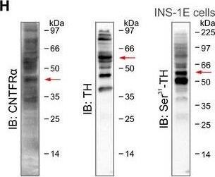
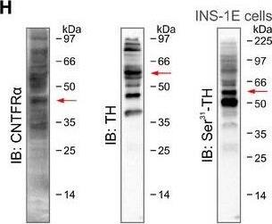
Secretagogin expression in rodent brain stem (related to Fig 4)A, A1Labeling specificity of our anti‐secretagogin antibody was verified in Scgn−/− mice (A1). These experiments also showed that tyrosine hydroxylase (TH)+ LC neurons (*) co‐express secretagogin in wild‐type (solid arrowheads) but not null mice (open arrowheads). Scale bars = 200 μm (low‐power micrographs, left) and 10 μm (high‐resolution insets, right).BSecretagogin+ neurons populate the locus coeruleus (LC) in the rat. Solid red circles pinpoint the general location of perikarya, while fine lines correspond to axons coursing toward the surface (4th ventricle).C, C1Secretagogin was expressed in both the somata (arrowheads; C) and dendrites (arrowheads; C1) of most TH+ neurons in the rat, too. Scale bars = 30 μm.DSecretagogin is localized to axons (arrowheads) in the rat. (D) Dorsal nucleus of vagus (nX), “2” shows the nucleus at high resolution. Scale bars = 200 μm (D), 30 μm (2).ESecretagogin (SCGN) is enriched in the synaptosomal fraction prepared from the mouse prefrontal cortex, lending additional support for its axonal localization. Synaptophysin (SYP) was used to control the enrichment of presynaptic proteins.FSecretagogin+ neurons can co‐express neuropeptide Y (NPY) in the LC of the rat. Open and solid arrowheads indicate the lack and existence of NPY co‐localization in adjacent neurons, respectively. Scale bars = 10 μm.GRepresentative Western blots from Cntf−/−, Scgn−/−, and wild‐type mice with or without intracerebroventricular CNTF treatment.H, H1INS‐1E cells were used as a cellular model exclusively because of their native co‐expression of CNTF receptor‐ alpha (CNTFR alpha), tyrosine hydroxylase (TH), secretagogin (SCGN), and extracellular signal‐regulated kinase 1/2 (Erk1/2). Red arrows label target proteins at their predicted sizes. Multiple bands likely represent splice variants, posttranslational modifications, or multimers. Antibodies were also validated histochemically often using knock‐out tissues.ISecretagogin, phospho‐Ser31‐TH, phospho‐Erk1, and total Erk1 expression in INS‐1E cells stimulated with CNTF with/without concomitant silencing of secretagogin (Scgn) gene expression (for 48 h). Note the lack of Erk1 phosphorylation upon genetic inactivation of secretagogin.JSignificant reduction of secretagogin (SCGN) protein levels upon acute RNAi‐mediated gene silencing in vitro (for 48 h). CNTF exposure did not affect the efficacy of gene silencing. Experiments were performed in triplicate; *P < 0.05 (Student's t‐test).KUnchanged tyrosine hydroxylase (TH) expression in wild‐type and Scgn−/− mice regardless of formalin‐induced stress (“+PFA”). These data validate our suggestion on protein phosphorylation, rather than degradation, being a critical step in secretagogin's mechanism of action. ns, non‐significant (Student's t‐test). Data are from n > 4 mice/genotype.Data information: Data are expressed as means ± s.e.m. Image collected and cropped by CiteAb from the following open publication (https://pubmed.ncbi.nlm.nih.gov/30209240), licensed under a CC-BY license. Not internally tested by R&D Systems.Applications for Rat CNTFR alpha Antibody
Immunohistochemistry
Sample: Perfusion fixed frozen sections of rat brain (cortex)
Western Blot
Sample: Recombinant Rat CNTF R alpha (Catalog # 558-CR)
Neutralization
Reviewed Applications
Read 2 reviews rated 5 using AF-559-NA in the following applications:
Formulation, Preparation, and Storage
Purification
Reconstitution
Formulation
Shipping
Stability & Storage
- 12 months from date of receipt, -20 to -70 °C as supplied.
- 1 month, 2 to 8 °C under sterile conditions after reconstitution.
- 6 months, -20 to -70 °C under sterile conditions after reconstitution.
Background: CNTFR alpha
The high-affinity CNTF receptor complex, which mediates the biological action of CNTF, contains three proteins: the ligand-binding a subunit (CNTF R alpha) and the two signal-transducing proteins LIF R beta and gp130. Whereas LIF R beta and gp130 are widely expressed in many cell types, the expression of CNTF R alpha is restricted to the central and peripheral nervous systems. cDNAs encoding CNTF R alpha have been isolated from both human and rat and were shown to share 94% amino acid (aa) sequence identity. Rat CNTF R alpha cDNA encodes a 372 amino acid residue precursor protein that apparently has a 22 aa residue signal peptide and five potential glycoslyation sites. CNTF R alpha differs from other cytokine receptors in that it lacks transmembrane and cytoplasmic domains and is anchored to cell membranes by a glycosylphosphatidylinositol (GPI) linkage. Similar to other GPI-linked proteins, soluble CNTF receptor alpha (CNTF sR alpha) can be released from the cell surface by phosphatidylinositol-specific phospholipase C. CNTF sR alpha can be released from skeletal muscle in response to peripheral nerve injury and high concentrations of CNTF sR alpha have also been detected in human cerebrospinal fluid. CNTF sR alpha binds CNTF in solution and the complex can act on cells that express only LIF R beta and gp130 but not CNTF R alpha.
References
- Yancopoulos, G.D. in Guidebook to Cytokines and Their Receptors, Nicola, N.A. editor, Oxford University Press, New York, pp137.
Long Name
Alternate Names
Gene Symbol
UniProt
Additional CNTFR alpha Products
Product Documents for Rat CNTFR alpha Antibody
Product Specific Notices for Rat CNTFR alpha Antibody
For research use only


