Rat Endoglin/CD105 Antibody
R&D Systems, part of Bio-Techne | Catalog # AF6440

Key Product Details
Species Reactivity
Validated:
Cited:
Applications
Validated:
Cited:
Label
Antibody Source
Product Specifications
Immunogen
Met1-Gly578
Accession # Q6Q3E8
Specificity
Clonality
Host
Isotype
Scientific Data Images for Rat Endoglin/CD105 Antibody
Detection of Rat Endoglin/CD105 by Western Blot.
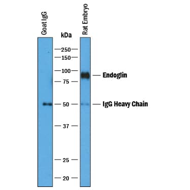
Western blot shows lysates of rat kidney tissue. PVDF membrane was probed with 1 µg/mL of Goat Anti-Rat Endoglin/CD105 Antigen Affinity-purified Polyclonal Antibody (Catalog # AF6440) followed by HRP-conjugated Anti-Goat IgG Secondary Antibody (Catalog # HAF017). A specific band was detected for Endoglin/CD105 at approximately 90 kDa (as indicated). This experiment was conducted under reducing conditions and using Immunoblot Buffer Group 1.Endoglin/CD105 in Rat Mesenchymal Stem Cells.
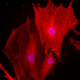
Endoglin/CD105 was detected in immersion fixed undifferentiated rat mesenchymal stem cells using Goat Anti-Rat Endoglin/CD105 Antigen Affinity-purified Polyclonal Antibody (Catalog # AF6440) at 10 µg/mL for 3 hours at room temperature. Cells were stained using the NorthernLights™ 557-conjugated Anti-Goat IgG Secondary Antibody (red; Catalog # NL001) and counterstained with DAPI (blue). Specific staining was localized to the transmembrane. View our protocol for Fluorescent ICC Staining of Stem Cells on Coverslips.Endoglin/CD105 in Rat Kidney.
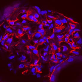
Endoglin/CD105 was detected in perfusion fixed frozen sections of rat kidney using Goat Anti-Rat Endoglin/CD105 Antigen Affinity-purified Polyclonal Antibody (Catalog # AF6440) at 1 µg/mL overnight at 4 °C. Tissue was stained using the NorthernLights™ 557-conjugated Anti-Goat IgG Secondary Antibody (red; Catalog # NL001) and counterstained with DAPI (blue). Specific staining was localized to endothelial cells in glomeruli. View our protocol for Fluorescent IHC Staining of Frozen Tissue Sections.Applications for Rat Endoglin/CD105 Antibody
Immunocytochemistry
Sample: Immersion fixed differentiated rat mesenchymal stem cells
Immunohistochemistry
Sample:
Perfusion fixed frozen sections of rat kidney.
Immunoprecipitation
Sample: Rat embryo tissue
Western Blot
Sample: Rat kidney tissue
Reviewed Applications
Read 1 review rated 4 using AF6440 in the following applications:
Formulation, Preparation, and Storage
Purification
Reconstitution
Formulation
Shipping
Stability & Storage
- 12 months from date of receipt, -20 to -70 °C as supplied.
- 1 month, 2 to 8 °C under sterile conditions after reconstitution.
- 6 months, -20 to -70 °C under sterile conditions after reconstitution.
Background: Endoglin/CD105
Endoglin (CD105) is a 90 kDa type I transmembrane glycoprotein of the zona pellucida (ZP) family of proteins. Endoglin and betaglycan/T betaRIII are type III receptors for TGF-beta superfamily ligands, sharing 71% aa identity with the transmembrane (TM) and cytoplasmic domains. Endoglin is highly expressed on proliferating vascular endothelial cells, chondrocytes, and syncytiotrophoblasts of term placenta, with lower amounts on hematopoietic, mesenchymal and neural crest stem cells, activated monocytes, and lymphoid and myeloid leukemic cells. Rat Endoglin cDNA encodes 650 amino acids (aa) including a 25 aa signal sequence, a 553 aa extracellular domain (ECD) with an orphan domain and a two-part ZP domain, a TM domain and a 51 aa cytoplasmic domain. In human and mouse, an isoform with a 14 aa cytoplasmic domain (S‑endoglin) can oppose effects of long (L) Endoglin. In rat, a potential isoform with a 100 aa cytoplasmic tail (49 aa inserted after aa 610) diverges at the same aa as S‑endoglin. The rat Endoglin ECD shares 84%, 70%, 68%, 64%, and 62% aa identity with mouse, human, canine, porcine, and bovine Endoglin, respectively. Endoglin homodimers interact with TGF-beta 1 and TGF-beta 3 (but not TGF-beta 2), but only after binding T betaRII. Similarly, they interact with activin-A and BMP-7 via activin type IIA or B receptors, and with BMP-2 via BMPR-1A/ALK-3 or BMPR-1B/ALK-6. BMP-9, however, is reported to bind Endoglin directly. Endoglin modifies ligand-induced signaling in multiple ways. For example, expression of Endoglin can inhibit TGF-beta 1 signals but enhance BMP7 signals in the same myoblast cell line. In endothelial cells, Endoglin inhibits T betaRI/ALK-5, but enhances ALK‑1‑mediated activation. Deletion of mouse Endoglin causes lethal vascular and cardiovascular defects, and human Endoglin haploinsufficiency can cause the vascular disorder, hereditary hemorrhagic telangiectasia type I. These abnormalities confirm the essential function of Endoglin in differentiation of smooth muscle, angiogenesis, and neovascularization. In preeclampsia of pregnancy, high levels of proteolytically generated soluble Endoglin and VEGF R1 (sFLT1), along with low placental growth factor (PlGF), are pathogenic due to anti-angiogenic activity.
Alternate Names
Gene Symbol
UniProt
Additional Endoglin/CD105 Products
Product Documents for Rat Endoglin/CD105 Antibody
Product Specific Notices for Rat Endoglin/CD105 Antibody
For research use only


