Rat Nogo-A aa 2-172 Antibody
R&D Systems, part of Bio-Techne | Catalog # AF3098

Key Product Details
Validated by
Biological Validation
Species Reactivity
Validated:
Rat
Cited:
Human, Mouse, Rat
Applications
Validated:
Immunohistochemistry, Western Blot
Cited:
Western Blot
Label
Unconjugated
Antibody Source
Polyclonal Goat IgG
Product Specifications
Immunogen
E. coli-derived recombinant rat Nogo-A
Glu2-Val172
Accession # Q9JK11
Glu2-Val172
Accession # Q9JK11
Specificity
Detects rat Nogo-A in direct ELISAs and Western blots.
Clonality
Polyclonal
Host
Goat
Isotype
IgG
Scientific Data Images for Rat Nogo-A aa 2-172 Antibody
Detection of Rat Nogo-A by Western Blot
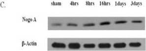
RT-PCR analysis and western blot analysis of Nogo-A mRNA and protein production in the hippocampus after TBI. (A) The PCR products from Nogo-A transcription after TBI with the yield of actin as an internal control. The sampling times after TBI are shown on the top (n = 4 in each group). (B) Quantification of Nogo-A mRNA expression by semiquantitative densitometry in conjunction with AlphaEase software (Alpha Innotech Corp.). (C) Time course of Nogo-A protein production after TBI, internal control: beta-actin. Times shown on top represent hours after injury. (D) Quantification of Nogo-A protein by semiquantitative densitometry in conjunction with AlphaEase software (Alpha Innotech Corp.). The data are presented as ratios related to the sham group. Bars represent the means ± SEM values. *p < 0.05 is considered significantly different from sham values using the Mann–Whitney U test. SEM, standard error of the mean; TBI, traumatic brain injury. Image collected and cropped by CiteAb from the following publication (https://jneuroinflammation.biomedcentral.com/articles/10.1186/1742-2094-9-121), licensed under a CC-BY license. Not internally tested by R&D Systems.Detection of Rat Nogo-A by Western Blot
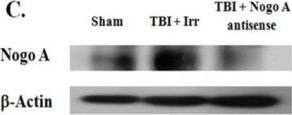
Effects of Nogo-A irrelevant control and antisense oligodeoxynucleotides on hippocampal Nogo-A expression after TBI. (A) RT-PCR analysis of Nogo-A mRNA transcription level. Actin transcription was used as an internal control. (B) The expression of Nogo-A was quantified by densitometry and compared with the data from rats injected with saline (sham), which was normalized to 100%. (C) Western blot analysis of Nogo-A protein level; beta-actin was used as an internal control. (D) Quantification of Nogo-A protein by semiquantitative densitometry in conjunction with AlphaEase software (Alpha Innotech Corp.). The data are presented compared with the sham group. The data are represented as the means ± SEM values (n = 6). *p < 0.05 was considered significantly different from the sham value using the Mann-Whiney U test, and #p < 0.05 was considered significantly different from the TBI with sense values using the Mann-Whiney U test. SEM, standard error of the mean; TBI, traumatic brain injury. Image collected and cropped by CiteAb from the following publication (https://jneuroinflammation.biomedcentral.com/articles/10.1186/1742-2094-9-121), licensed under a CC-BY license. Not internally tested by R&D Systems.Detection of Rat Nogo-A by Western Blot
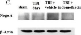
Effects of indomethacin administration on Nogo-A expression. Animals were in one of four groups: sham (no TBI), TBI treatment (TBI eight hours), TBI combined with vehicle administration (TBI + vehicle), and TBI combined with indomethacin administration (TBI + indomethacin). (A) RT-PCR analysis of the expression of Nogo-A among different groups along with the analysis of beta-actin transcription as an internal control. (B) Quantification of Nogo-A expression. (C) Western blot analysis of the expression of Nogo-A among different groups along with the analysis of beta-actin as an internal control. (D) Quantification of Nogo-A expression. Bars represent means ± SEM values (n = 5). *P <0.05 is considered significantly different from the sham value using the Mann–Whitney U test and #P <0.05 is considered significantly different from the TBI value using the Mann–Whitney U test. SEM, standard error of the mean; TBI, traumatic brain injury. Image collected and cropped by CiteAb from the following publication (https://jneuroinflammation.biomedcentral.com/articles/10.1186/1742-2094-9-121), licensed under a CC-BY license. Not internally tested by R&D Systems.Applications for Rat Nogo-A aa 2-172 Antibody
Application
Recommended Usage
Immunohistochemistry
5-15 µg/mL
Sample: Perfusion fixed frozen sections of rat brain (caudate putamen)
Sample: Perfusion fixed frozen sections of rat brain (caudate putamen)
Western Blot
0.1 µg/mL
Sample: Recombinant Rat Nogo-A Fc Chimera (Catalog # 2445-NG)
Sample: Recombinant Rat Nogo-A Fc Chimera (Catalog # 2445-NG)
Formulation, Preparation, and Storage
Purification
Antigen Affinity-purified
Reconstitution
Reconstitute at 0.2 mg/mL in sterile PBS. For liquid material, refer to CoA for concentration.
Formulation
Lyophilized from a 0.2 μm filtered solution in PBS with Trehalose. *Small pack size (SP) is supplied either lyophilized or as a 0.2 µm filtered solution in PBS.
Shipping
Lyophilized product is shipped at ambient temperature. Liquid small pack size (-SP) is shipped with polar packs. Upon receipt, store immediately at the temperature recommended below.
Stability & Storage
Use a manual defrost freezer and avoid repeated freeze-thaw cycles.
- 12 months from date of receipt, -20 to -70 °C as supplied.
- 1 month, 2 to 8 °C under sterile conditions after reconstitution.
- 6 months, -20 to -70 °C under sterile conditions after reconstitution.
Background: Nogo-A
Long Name
Reticulon 4A
Alternate Names
NI220, NogoA, RTN4, RTN4A
Gene Symbol
RTN4
UniProt
Additional Nogo-A Products
Product Documents for Rat Nogo-A aa 2-172 Antibody
Product Specific Notices for Rat Nogo-A aa 2-172 Antibody
For research use only
Loading...
Loading...
Loading...
Loading...


