Tau [p Thr205, p Ser202] Antibody (AH36) - BSA Free
Novus Biologicals, part of Bio-Techne | Catalog # NBP3-18265
Recombinant Monoclonal Antibody


Conjugate
Catalog #
Key Product Details
Species Reactivity
Human, Mouse
Applications
Dot Blot, Immunocytochemistry/ Immunofluorescence, Immunohistochemistry, Western Blot
Label
Unconjugated
Antibody Source
Monoclonal Rabbit IgG Clone # AH36
Format
BSA Free
Concentration
1 mg/ml
Product Specifications
Immunogen
Synthetic peptide of Human Phospho Tau (Ser202/Thr205)
Modification
p Ser 202, p Thr 205
Localization
Cytoskeleton|Cytosol|Plasma Membrane|Axon|Dendrite
Clonality
Monoclonal
Host
Rabbit
Isotype
IgG
Scientific Data Images for Tau [p Thr205, p Ser202] Antibody (AH36) - BSA Free
Western Blot: Tau [p Ser 202, p Thr 205] Antibody (AH36) [NBP3-18265] - Western Blot analysis of Human iPSC-derived cortical neurons showing detection of Tau protein using Rabbit Anti-Tau Monoclonal Antibody, Clone AH36 (NBP3-18265). Lane 1: MW ladder. Lane 2: Control (non-disease) line. Lane 2: Ex10+16 tau mutant sample. Lane 3: P301L tau mutant sample. Load: 50ug. Primary Antibody: Rabbit Anti-Tau Monoclonal Antibody (NBP3-18265) at 1:500 for Overnight. pSer202/pThr205 was detected using NBP3-18265. Total tau was detected using mouse anti-tau antibody (clone HT7). The bar graph on the right shows quantification of pSer202/pThr205 compared to total tau in each sample.
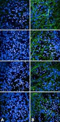
Immunocytochemistry/Immunofluorescence: Tau [p Ser 202, p Thr 205] Antibody (AH36) [NBP3-18265] - Immunocytochemistry/Immunofluorescence analysis using Rabbit Anti-Tau Monoclonal Antibody, Clone AH36 (NBP3-18265). Tissue: iPSC-derived cortical excitatory neurons. Species: Human. Primary Antibody: Rabbit Anti-Tau Monoclonal Antibody (NBP3-18265) at 1:500 for Overnight. Secondary Antibody: Donkey anti-rabbit: Alexa Fluor 488 at 1:1000. Counterstain: DAPI. A) iPSC-derived neurons from non-demented control (NDC). B) iPSC-derived neurons from subject with P301L MAPT mutation. Images acquired using an automated Opera Phoenix system. Each field of view is a max projection from 10 planes of 1 um stacks.
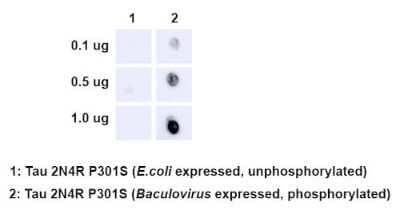
Dot Blot: Tau [p Ser 202, p Thr 205] Antibody (AH36) [NBP3-18265] - Dot Blot analysis using Rabbit Anti-Tau Monoclonal Antibody, Clone AH36 (NBP3-18265). Species: E. Coli, Baculovirus. Primary Antibody: Rabbit Anti-Tau Monoclonal Antibody (NBP3-18265) at 1:500. Secondary Antibody: Goat anti-rabbit IgG:HRP.
Applications for Tau [p Thr205, p Ser202] Antibody (AH36) - BSA Free
Application
Recommended Usage
Immunocytochemistry/ Immunofluorescence
1:500
Immunohistochemistry
1:500
Western Blot
1:500
Application Notes
Optimal dilutions for assays should be determined by the user.
Formulation, Preparation, and Storage
Purification
Affinity purified
Formulation
PBS pH 7.4, 50% glycerol
Format
BSA Free
Preservative
0.09% Sodium Azide
Concentration
1 mg/ml
Shipping
The product is shipped with polar packs. Upon receipt, store it immediately at the temperature recommended below.
Stability & Storage
Store at -20C. Avoid freeze-thaw cycles.
Background: Tau
Long Name
Microtubule-Associated Protein Tau
Alternate Names
MAPT, MSTD, MTBT1, Neurofibrillary tangle protein, PPND
Gene Symbol
MAPT
Additional Tau Products
Product Documents for Tau [p Thr205, p Ser202] Antibody (AH36) - BSA Free
Product Specific Notices for Tau [p Thr205, p Ser202] Antibody (AH36) - BSA Free
This product is for research use only and is not approved for use in humans or in clinical diagnosis. Primary Antibodies are guaranteed for 1 year from date of receipt.
Loading...
Loading...
Loading...
Loading...

