Human Pluripotent Stem Cell Marker Antibody Panel
R&D Systems, part of Bio-Techne | Catalog # SC008

Key Product Details
Assay Procedure
Refer to the product datasheet for complete product details.
Briefly, human stem cell pluripotency can be verified using marker antibodies and the following procedure:
- The cells are harvested and processed for either immunocytochemistry or flow cytometry
- The cells are incubated with antibody markers of pluripotency
- Pluripotency marker expression is analyzed
Reagents supplied in the Human Pluripotent Stem Cell Marker Antibody Panel Kit (Catalog # SC008):
Positive Markers
- Mouse Anti-Human Alkaline Phosphatase Monoclonal Antibody
- Goat Anti-Human Nanog Antigen-affinity Purified Polyclonal Antibody
- Goat Anti-Human Oct-3/4 Antigen-affinity Purified Polyclonal Antibody
- Mouse Anti-Human SSEA-4 Monoclonal Antibody
Negative Markers
- Mouse Anti-Human SSEA-1 Monoclonal Antibody
Immunocytochemistry
Reagents
- Appropriate stem cell culture substrate (e.g., StemXVivo® Culture Matrix (Catalog # NL001), iMEFs (Catalog # PSC001), etc.)
- Cell culture medium
- Sterile PBS
- 4% paraformaldehyde in PBS
- 1% BSA in PBS
- 0.1% Triton™ X-100, 1% BSA, 10% normal donkey serum in PBS
- Mounting medium (Catalog # CTS011 or equivalent)
- Secondary developing reagents (Catalog # NL001, NL003, NL007, NL009, or equivalent)
Materials
- Human pluripotent stem cells
- Cell culture plate (24-well)
Equipment
- 37 °C, 5% CO2 incubator
- Centrifuge
- Hemocytometer
- Inverted microscope
- Fluorescence microscope
Flow Cytometry
Reagents
- Isotype controls (Catalog # MAB002 and MAB007, or equivalent)
- FACS buffer (2% fetal bovine serum, 0.1% sodium azide in Hank’s buffer)
- Secondary developing reagents
Materials
- Human pluripotent stem cells
- 5 mL tubes
Equipment
- 37 °C, 5% CO2 incubator
- 2 °C to 8 °C refrigerator
- Centrifuge
- Hemocytometer
- Flow cytometer
Triton is a registered trademark of Union Carbide, Inc.
Immunocytochemistry
Pass a suspension of mouse bone marrow cells through a 70 μm nylon strainer to remove clumps and debris.
Remove red blood cells if necessary.
Wash the cells with IMDM/2% FBS by centrifugation at 400 x g for 10 minutes and pool the cells.

Coat coverslips with stem cell subtype-specific substrate.

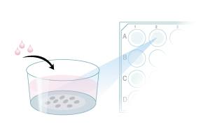
Plate stem cells.
Culture to desired density/age.

Fix stem cells with 4% paraformaldehyde.

Block with blocking solution.

Incubate with primary antibodies.
Wash with wash buffer.

Incubate with fluorochrome-conjugated secondary antibodies.
Wash with wash buffer.

Incubate with nuclear counterstain.

Mount the coverslip.
Visualize using a fluorescence microscope and appropriate filter sets.

Flow Cytometry
Perform a cell count on harvested cells.
Resuspend the cells in FACS buffer at 1 x 10⁶ cells/mL.

Aliquot 90 µL of the cell suspension into a 5 mL flow cytometry tube.

Add 10 µL of each antibody or isotype control to the cells.
Incubate for 30 minutes at room temperature.

Centrifuge samples at 300 x g for 5 minutes.
Wash the cells three times with FACS buffer.
Resuspend the cells in 200 µL FACS buffer.

Add 10 µL of a fluorochrome-conjugated secondary developing reagent.
Incubate for 30 minutes at room temperature in the dark.

Centrifuge samples at 300 x g for 5 minutes.
Wash the cells three times with FACS buffer.
Resuspend the cells in 200 µL FACS buffer.

Analyze the cells by flow cytometry.

Customer Reviews for Human Pluripotent Stem Cell Marker Antibody Panel
There are currently no reviews for this product. Be the first to review Human Pluripotent Stem Cell Marker Antibody Panel and earn rewards!
Have you used Human Pluripotent Stem Cell Marker Antibody Panel?
Submit a review and receive an Amazon gift card!
$25/€18/£15/$25CAN/¥2500 Yen for a review with an image
$10/€7/£6/$10CAN/¥1110 Yen for a review without an image
Submit a review


