Product Overview
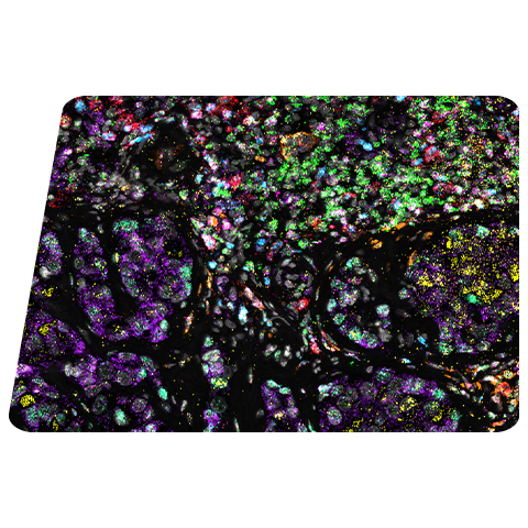
RNAscope™ HiPlex assay can be combined with Imaging Mass Cytometry™ (IMC™) to enable same-slide co-detection of RNA and protein targets on FFPE tissues.
IMC is a proven tool for the study of complex cellular interactions in the TME. It utilizes CyTOF technology for simultaneous assessment of 40-plus protein markers at subcellular resolution without spectral overlap or background autofluorescence, thus providing unprecedented insight into the tissue organization and function. The RNAscope HiPlex v2 assay can detect up to 12 targets on FFPE tissues. By using metal-conjugated instead of fluorophore-conjugated labeled probes, RNA targets can be detected using the Hyperion Imaging Mass Cytometry system.
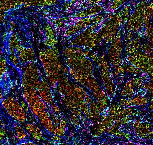
Designed for Imaging Mass Cytometry™-based Hyperion Imaging Systems
- No interference from autofluorescence
- Simultaneous imaging of 40 markers
RNA and Protein Co-detection
- Stain up to 12 RNA targets using RNAscope HiPlex probes.
- Utilize remaining metals to stain proteins.
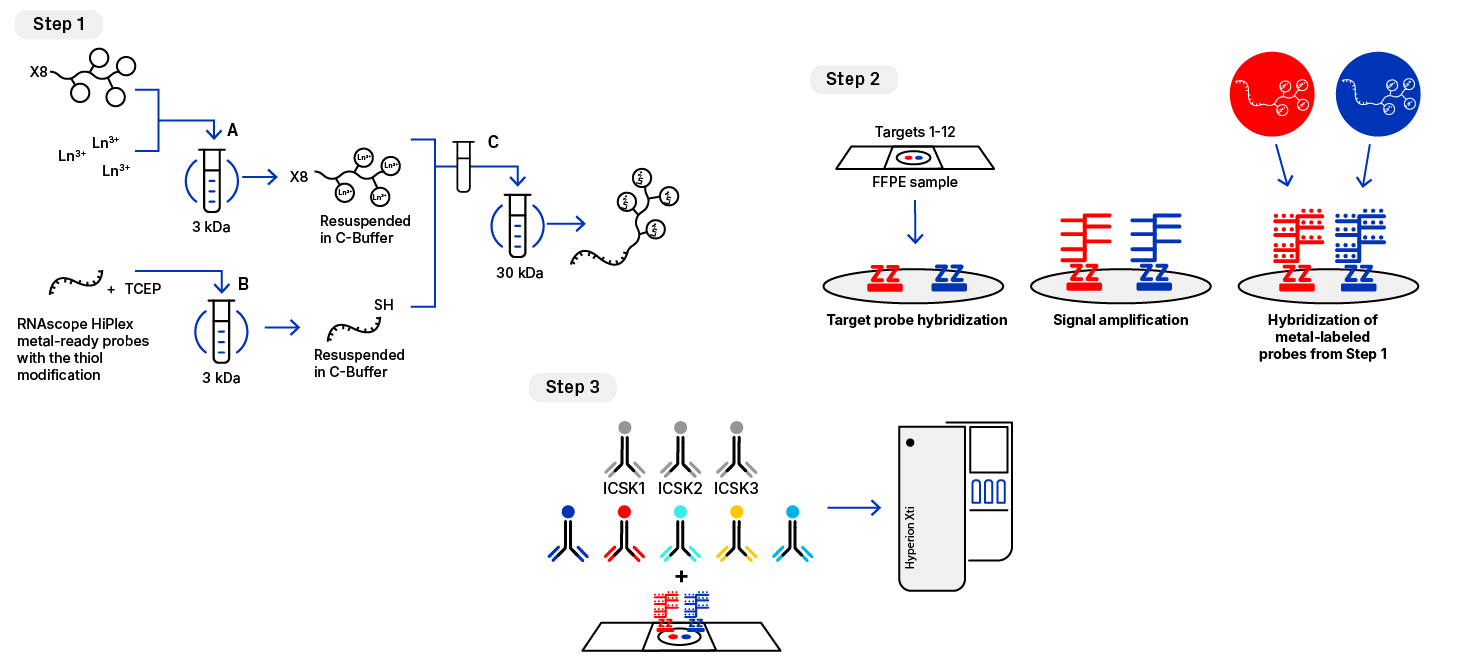
Figure 1. Schematic showing the 3-step procedure to acquire RNA and protein co-detection data with IMC. Step 1 - Labeling of detection oligonucleotides. (A) Preloading the X8 polymer lanthanide metal, (B) Reduction of the metal-ready probe, (C) Combination, conjugation and purification with spin filter; Step 2- Modified RNAscope Hiplex Flex Assay v2; Step 3 - IMC antibody staining overnight.
Our technical note provides a workflow and procedure to achieve RNA and protein co-detection data in IMC experiments. This workflow utilizes the highly sensitive and specific RNAscope technology for RNA detection with the multiplexing capability of IMC to visualize key RNA and protein markers in the same FFPE samples.
Immuno-oncology
- Detect immune cells using marker antibodies.
- Visualize cytokines and chemokines using RNAscope probes and determine immune activation states.
Immunology
Investigate immune response to disease, infection or treatment.
Find RNAscope Probes
Find probes below or request a custom design if you don’t see your target.
Ordering Information
Refer to the Technical Note for a complete list of additional materials and equipment required for the procedure.
| Labeling Kit (Standard Biotools) | Product Information |
|---|---|
| Maxpar X8 Antibody labeling kit | Select the metal-specific kit of your choice |
| Reagent Kit (ACD, Bio-Techne) | Catalog # |
|---|---|
| RNAscope™ HiPlex12 Flex Reagents Kit v2 (without Fluoros) | 322725 |
| RNAscope HiPlex12 Reagents Kit v2 (with Fluoros) | 324409 |
