Multiomic Mapping of the Brain: Same-Section, Fully-Automated Spatial RNA and Protein Detection on Mouse Frozen Tissues
Scientific Meeting Posters
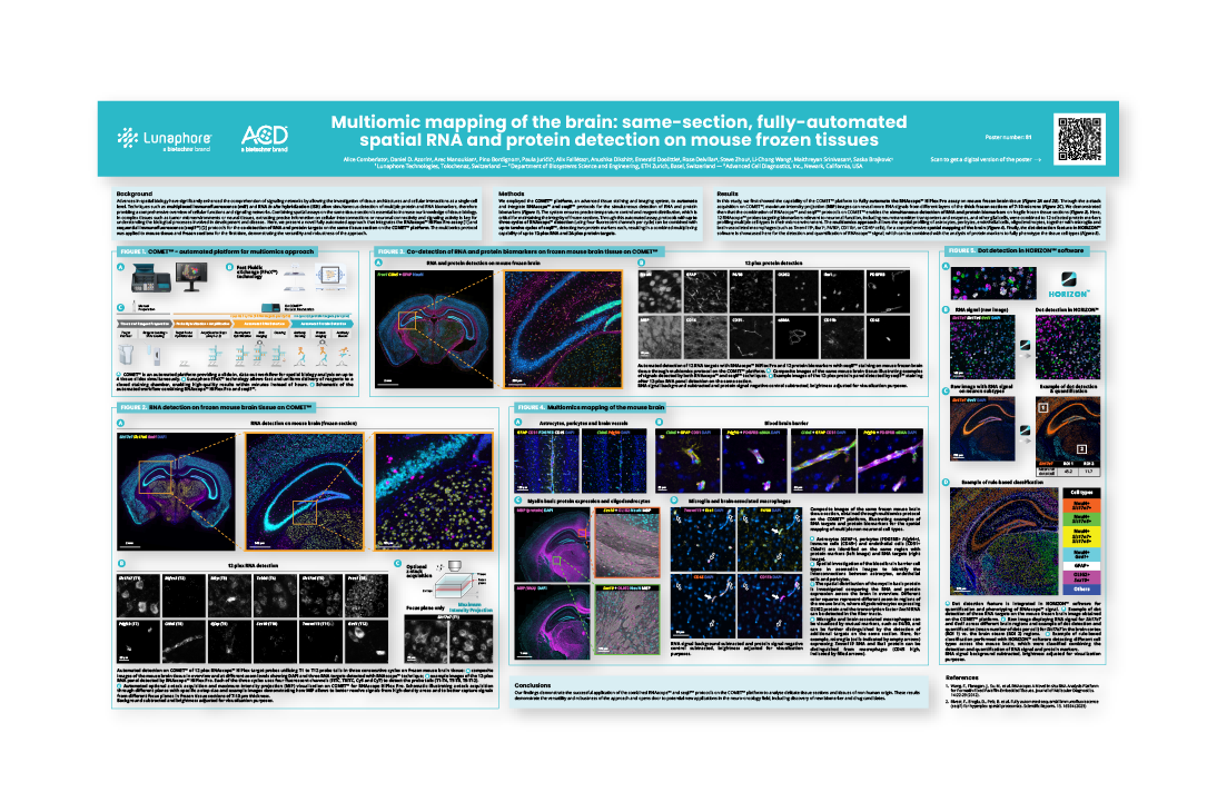
Advances in spatial biology have significantly enhanced the comprehension of signaling networks by allowing the investigation of tissue architectures and cellular interactions at a single-cell level. Techniques such as multiplexed immunofluorescence (mIF) and RNA in situ hybridization (ISH) allow simultaneous detection of multiple protein and RNA biomarkers, therefore providing a comprehensive overview of cellular functions and signaling networks. Combining spatial assays on the same tissue section is essential to increase our knowledge of tissue biology.
In complex tissues such as tumor microenvironments or neural tissues, extracting precise information on cellular interconnections or neuronal connectivity and signaling activity is key for understanding the biological processes involved in development and disease. Here, we present a novel fully automated approach that integrates the RNAscope™ HiPlex Pro assay [1] and sequential immunofluorescence (seqIF™) [2] protocols for the co-detection of RNA and protein targets on the same tissue section on the COMET™ platform. The multiomics protocol was applied to mouse tissue and frozen sections for the first time, demonstrating the versatility and robustness of the approach.