Equine IL-10 Antibody
R&D Systems, part of Bio-Techne | Catalog # AF1605


Key Product Details
Validated by
Species Reactivity
Validated:
Cited:
Applications
Validated:
Cited:
Label
Antibody Source
Product Specifications
Immunogen
Ser19-Asn178
Accession # Q28374
Specificity
Clonality
Host
Isotype
Endotoxin Level
Scientific Data Images for Equine IL-10 Antibody
Cell Proliferation Induced by IL‑10 and Neutralization by Equine IL‑10 Antibody.
Recombinant Equine IL-10 (Catalog # 1605-IL) stimulates proliferation in the MC/9-2 mouse mast cell line in a dose-dependent manner (orange line). Proliferation elicited by Recombinant Equine IL-10 (20 ng/mL) is neutralized (green line) by increasing concentrations of Goat Anti-Equine IL-10 Antigen Affinity-purified Polyclonal Antibody (Catalog # AF1605). The ND50 is typically 0.2-0.6 µg/mL.IL‑10 in Equine PBMCs.
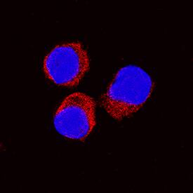
IL-10 was detected in immersion fixed equine peripheral blood mononuclear cells (PBMCs) treated with calcium ionomycin and PMA using Goat Anti-Equine IL-10 Antigen Affinity-purified Polyclonal Antibody (Catalog # AF1605) at 15 µg/mL for 3 hours at room temperature. Cells were stained using the NorthernLights™ 557-conjugated Anti-Goat IgG Secondary Antibody (red; Catalog # NL001) and counterstained with DAPI (blue). Specific staining was localized to cytoplasm. View our protocol for Fluorescent ICC Staining of Non-adherent Cells.Applications for Equine IL-10 Antibody
Immunocytochemistry
Sample: Immersion fixed equine peripheral blood mononuclear cells
Western Blot
Sample: Recombinant Equine IL-10 (Catalog # 1605-IL)
Neutralization
Equine IL-10 Sandwich Immunoassay
Formulation, Preparation, and Storage
Purification
Reconstitution
Formulation
Shipping
Stability & Storage
- 12 months from date of receipt, -20 to -70 °C as supplied.
- 1 month, 2 to 8 °C under sterile conditions after reconstitution.
- 6 months, -20 to -70 °C under sterile conditions after reconstitution.
Background: IL-10
Interleukin 10 (IL-10), initially designated cytokine synthesis inhibitory factor (CSIF), was originally identified as a product of mouse T helper 2 (Th2) cells that inhibited the cytokine production by Th1 cells. It is a pleiotropic cytokine that regulates the immune and inflammatory responses of hematopoietic cells (1, 2). IL-10 has immunosuppressive activities and has been shown to inhibit the effector functions of monocyte/macrophage and CD4+ T cells. Conversely, IL-10 has immunostimulatory activities and can induce the proliferation and cytotoxic activity of CD8+ T cells and NK cells. IL-10 also regulates the growth and differentiation of B cells, mast cells, dendritic cells and neutrophils (1). The biological activities of IL-10 is mediated by the heteromeric IL-10 receptor complex, which is composed of the ligand-binding IL-10R alpha and the accessory IL-10R beta subunits. Both subunits belong to the class II cytokine receptor family. IL-10R beta is also utilized as a subunit in the heterodimer receptor complex for IL-22, IL-28 and IL-29. Besides IL-10, five novel cytokines (IL-19, -20, -22, -24, and -26) that share structural and limited sequence homology with IL-10 have been identified. These proteins constitute the IL-10 cytokine family (3).
Equine IL-10 cDNA encodes a 178 amino acid residue (aa) precursor protein with an 18 aa signal peptide and 160 aa mature protein that contains two potential N-linked glycosylation sites. Analogous to human IL-10, equine IL-10 likely exists as nondisulfide-linked homodimers. Equine IL-10 shares 71% and 78% aa sequence homology with mouse and human IL-10, respectively.
References
- Moore, K. et al. (2001) Annu. Rev. Immunol. 19:683.
- Mocellin, S. et al. (2003) Trends in Immunol. 23:36.
- Conti, P. et al. (2003) Immunol. Letters 88:171.
Long Name
Alternate Names
Entrez Gene IDs
Gene Symbol
UniProt
Additional IL-10 Products
Product Documents for Equine IL-10 Antibody
Product Specific Notices for Equine IL-10 Antibody
For research use only
