Human ABCG2 PE-conjugated Antibody
R&D Systems, part of Bio-Techne | Catalog # FAB995P


Conjugate
Catalog #
Key Product Details
Species Reactivity
Validated:
Human
Cited:
Human
Applications
Validated:
Flow Cytometry
Cited:
Flow Cytometry
Label
Phycoerythrin (Excitation = 488 nm, Emission = 565-605 nm)
Antibody Source
Monoclonal Mouse IgG2B Clone # 5D3
Product Specifications
Immunogen
3T3 cells transduced with human ABCG2
Specificity
Detects human ABCG2 in flow cytometry and immunocytochemistry.
Clonality
Monoclonal
Host
Mouse
Isotype
IgG2B
Scientific Data Images for Human ABCG2 PE-conjugated Antibody
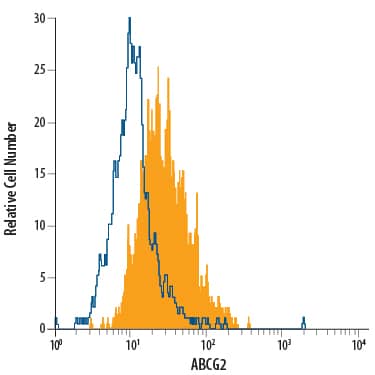
Detection of ABCG2 in MCF‑7 Human Cell Line by Flow Cytometry.
MCF-7 human breast cancer cell line was stained with Mouse Anti-Human ABCG2 PE-conjugated Monoclonal Antibody (Catalog # FAB995P, filled histogram) or isotype control antibody (IC0041P, open histogram). View our protocol for Staining Membrane-associated Proteins.Detection of Porcine ABCG2 by Flow Cytometry
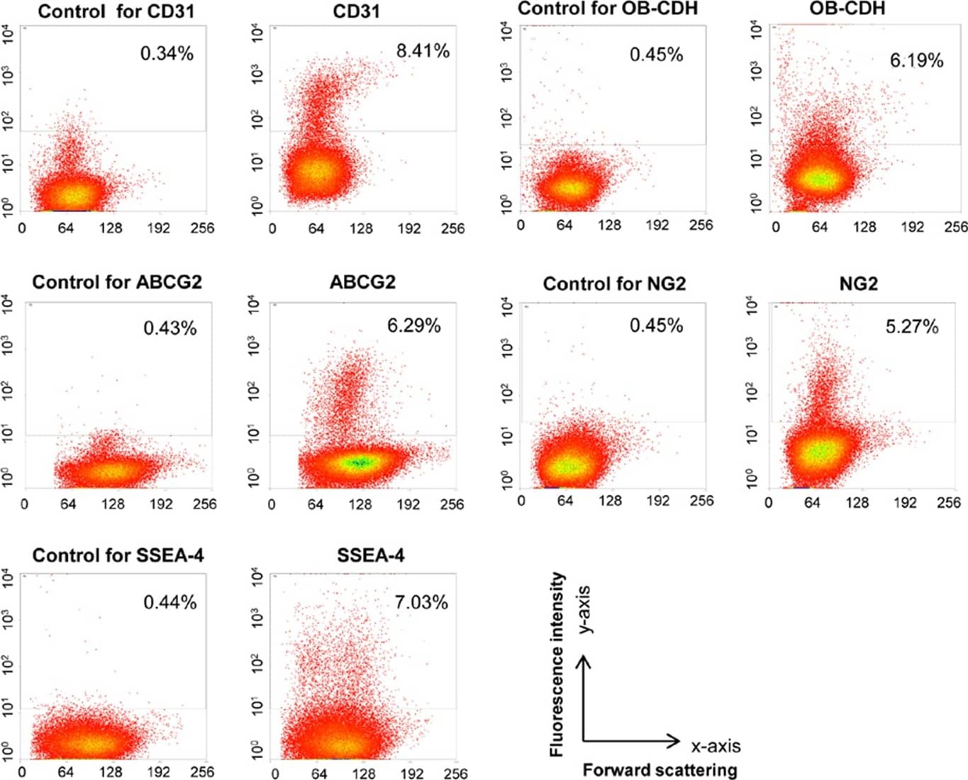
Freshly isolated aortic pVICs express distinct cell surface markers.Freshly isolated aortic pVICs were stained with antibodies for CD31, OB–CDH, ABCG2, NG2, SSEA-4, and the corresponding control antibodies. Staining was quantified by flow cytometry. In the figure, the y-axis is fluorescence intensity and the x-axis is forward scattering. Percentage in the rectangular gates represents the fraction of positively stained cells. After subtracting the background, about 7.70% of these aortic pVICs stained positive for CD31, 4.71% stained positive for OB–CDH, 5.60% stained positive for ABCG2, 5.56% stained positive for NG2, and 6.59% stained positive for SSEA-4. Image collected and cropped by CiteAb from the following publication (https://dx.plos.org/10.1371/journal.pone.0069667), licensed under a CC-BY license. Not internally tested by R&D Systems.Detection of ABCG2 in RPMI 8226 cells by Flow Cytometry
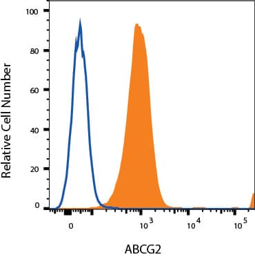
RPMI 8226 cells were stained with Mouse Anti-Human ABCG2 PE-conjugated Monoclonal Antibody (Catalog # FAB995P, filled histogram) or isotype control antibody (Catalog # IC0041P, open histogram). View our protocol for Staining Membrane-associated Proteins.Applications for Human ABCG2 PE-conjugated Antibody
Application
Recommended Usage
Flow Cytometry
10 µL/106 cells
Sample: MCF‑7 human breast cancer cell line; RPMI 8226 human multiple myeloma cell line
Sample: MCF‑7 human breast cancer cell line; RPMI 8226 human multiple myeloma cell line
Reviewed Applications
Read 1 review rated 4 using FAB995P in the following applications:
Formulation, Preparation, and Storage
Purification
Protein A or G purified from hybridoma culture supernatant
Formulation
Supplied in a saline solution containing BSA and Sodium Azide.
Shipping
The product is shipped with polar packs. Upon receipt, store it immediately at the temperature recommended below.
Stability & Storage
Protect from light. Do not freeze.
- 12 months from date of receipt, 2 to 8 °C as supplied.
Background: ABCG2
References
- Chaudhary, P.M. and I.B. Roninson (1991) Cell 66:85.
- Sorrentino, B.P. et al. (1995) Blood 86:491.
- Pallis, M. and N. Russell (2000) Blood 95:2897.
- Johnstone, R.W. et al. (1999) Blood 93:1075.
- Doyle, L.A. et al. (1998) Proc. Natl. Acad. Sci. USA 95:15665.
- Zhou, S. et al. (2001) Nat. Medicine 7:1028.
- Hrycyna, C.A. et al. (1998) Biochem. 37:13660.
- Bunting, K.D. (2002) Stem Cells 20:11.
Long Name
ATP-binding Cassette Transporter G2
Alternate Names
ABCP, BCRP, CD338, MXR
Entrez Gene IDs
9429 (Human)
Gene Symbol
ABCG2
Additional ABCG2 Products
Product Documents for Human ABCG2 PE-conjugated Antibody
Product Specific Notices for Human ABCG2 PE-conjugated Antibody
For research use only
Loading...
Loading...
Loading...
Loading...
Loading...

