Human CCL2/JE/MCP-1 Antibody
R&D Systems, part of Bio-Techne | Catalog # MAB679

Key Product Details
Validated by
Biological Validation
Species Reactivity
Validated:
Human
Cited:
Human, Mouse, Porcine
Applications
Validated:
Dual RNAscope ISH-IHC Compatible, ELISA Capture (Matched Antibody Pair), Immunohistochemistry, Neutralization, Western Blot
Cited:
Antibody Array Development, Bioassay, ELISA, ELISA Capture, ELISA Development, ELISA Development (Capture), Functional Assay, Immunocytochemistry, Immunohistochemistry-Paraffin, Luminex Development, Neutralization, Western Blot
Label
Unconjugated
Antibody Source
Monoclonal Mouse IgG2B Clone # 23007
Product Specifications
Immunogen
E. coli-derived recombinant human CCL2/JE/MCP-1
Specificity
Detects human CCL2/JE/MCP-1 in ELISAs and Western blots. In ELISAs, this antibody does not cross-react with recombinant mouse (rm) CCL2, 3, 4, rhCCL3, 4, 5, 7, or 8.
Clonality
Monoclonal
Host
Mouse
Isotype
IgG2B
Endotoxin Level
<0.10 EU per 1 μg of the antibody by the LAL method.
Scientific Data Images for Human CCL2/JE/MCP-1 Antibody
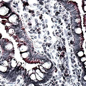
CCL2/JE/MCP‑1 in Human Crohn's Disease Intestine.
CCL2/JE/MCP-1 was detected in immersion fixed paraffin-embedded sections of human Crohn's disease intestine using Mouse Anti-Human CCL2/JE/MCP-1 Monoclonal Antibody (Catalog # MAB679) at 15 µg/mL for 1 hour at room temperature followed by incubation with the Anti-Mouse IgG VisUCyte™ HRP Polymer Antibody (Catalog # VC001). Before incubation with the primary antibody, tissue was subjected to heat-induced epitope retrieval using Antigen Retrieval Reagent-Basic (Catalog # CTS013). Tissue was stained using DAB (brown) and counterstained with hematoxylin (blue). Specific staining was localized to cell membranes and cytoplasm. View our protocol for IHC Staining with VisUCyte HRP Polymer Detection Reagents.Chemotaxis Induced by CCL2/MCP-1 and Neutralization by Human CCL2/MCP‑1 Antibody.
Recombinant Human CCL2/MCP-1 (Catalog # 279-MC) chemoattracts the BaF3 mouse pro-B cell line transfected with human CCR2A in a dose-dependent manner (orange line). The amount of cells that migrated through to the lower chemotaxis chamber was measured by Resazurin (Catalog # AR002). Chemotaxis elicited by Recombinant Human CCL2/MCP-1 (75 ng/mL) is neutralized (green line) by increasing concentrations of Human CCL2/MCP-1 Monoclonal Antibody (Catalog # MAB679). The ND50 is typically 0.5-2.0 µg/mL.Detection of Human CCL2/JE/MCP-1 by Immunohistochemistry
Expression patterns of CCL2, CCL5, TNF alpha and IL-1 beta in healthy individuals and breast cancer patients. Representative examples of the expression of CCL2, CCL5, TNF alpha and IL-1 beta in the different groups of patients included in the study, in biopsies obtained at the time of diagnosis. (a1-a4) Patients diagnosed with benign breast disorders. The pictures demonstrate the lack of staining of the four factors in the normal breast epithelial cells, as denoted in the majority of patients included in this group. (b1-b4) DCIS patients. The pictures demonstrate positive staining of the four factors in the malignant lesions, as denoted in the majority of patients included in this group. (c1-c4) IDC-no-relapse patients. The pictures demonstrate positive staining of the four factors in the tumor cells, as denoted in the majority of patients included in this group. (d1-d4) IDC-with-relapse patients. The pictures demonstrate positive staining of the four factors in the tumor cells, as denoted in the majority of patients included in this group. (a1, b1, c1, d1) CCL2 staining; (a2, b2, c2, d2) CCL5 staining; (a3, b3, c3, d3) TNF alpha staining; (a4, b4, c4, d4) IL-1 beta staining. The expression of the proteins was determined by IHC using specific antibodies, whose specificity in IHC was verified. The values of photo magnification are indicated in the left bottom corner of each of the pictures. Image collected and cropped by CiteAb from the following publication (https://pubmed.ncbi.nlm.nih.gov/21486440), licensed under a CC-BY license. Not internally tested by R&D Systems.Applications for Human CCL2/JE/MCP-1 Antibody
Application
Recommended Usage
Dual RNAscope ISH-IHC Compatible
5-15 µg/mL
Sample: Immersion fixed paraffin-embedded sections of human Crohn's disease
Sample: Immersion fixed paraffin-embedded sections of human Crohn's disease
Immunohistochemistry
5-15 µg/mL
Sample: Immersion fixed paraffin-embedded sections of human Crohn's disease intestine
Sample: Immersion fixed paraffin-embedded sections of human Crohn's disease intestine
Western Blot
1 µg/mL
Sample: Recombinant Human CCL2/JE/MCP-1 (Catalog # 279-MC)
Sample: Recombinant Human CCL2/JE/MCP-1 (Catalog # 279-MC)
Neutralization
Measured by its ability to neutralize CCL2/JE/MCP‑1-induced chemotaxis in the BaF3 mouse pro‑B cell line transfected with human CCR2A. The Neutralization Dose (ND50) is typically 0.5-2.0 µg/mL in the presence of 75 ng/mL Recombinant Human CCL2/JE/MCP‑1.
Human CCL2/JE/MCP-1 Sandwich Immunoassay
Please Note: Optimal dilutions of this antibody should be experimentally determined.
Reviewed Applications
Read 3 reviews rated 5 using MAB679 in the following applications:
Formulation, Preparation, and Storage
Purification
Protein A or G purified from ascites
Reconstitution
Reconstitute at 0.5 mg/mL in sterile PBS. For liquid material, refer to CoA for concentration.
Formulation
Lyophilized from a 0.2 μm filtered solution in PBS with Trehalose. See Certificate of Analysis for details.
*Small pack size (-SP) is supplied either lyophilized or as a 0.2 µm filtered solution in PBS.
*Small pack size (-SP) is supplied either lyophilized or as a 0.2 µm filtered solution in PBS.
Shipping
Lyophilized product is shipped at ambient temperature. Liquid small pack size (-SP) is shipped with polar packs. Upon receipt, store immediately at the temperature recommended below.
Stability & Storage
Use a manual defrost freezer and avoid repeated freeze-thaw cycles.
- 12 months from date of receipt, -20 to -70 °C as supplied.
- 1 month, 2 to 8 °C under sterile conditions after reconstitution.
- 6 months, -20 to -70 °C under sterile conditions after reconstitution.
Background: CCL2/JE/MCP-1
Alternate Names
GDCF-2, HC11, HSMCR30, MCAF, MCP-1, SMC-CF
Gene Symbol
CCL2
Additional CCL2/JE/MCP-1 Products
Product Documents for Human CCL2/JE/MCP-1 Antibody
Product Specific Notices for Human CCL2/JE/MCP-1 Antibody
For research use only
Loading...
Loading...
Loading...
Loading...


