Human CD160 Antibody
R&D Systems, part of Bio-Techne | Catalog # MAB6700


Conjugate
Catalog #
Key Product Details
Species Reactivity
Validated:
Human
Cited:
Human
Applications
Validated:
CyTOF-ready, Flow Cytometry
Cited:
Functional Assay, Neutralization
Label
Unconjugated
Antibody Source
Monoclonal Mouse IgG2B Clone # 688327
Product Specifications
Immunogen
Chinese hamster ovary cell line CHO-derived recombinant human CD160
Ile27-Ser159
Accession # O95971
Ile27-Ser159
Accession # O95971
Specificity
Detects human CD160 in direct ELISAs.
In direct ELISAs and Western blots, no
cross-reactivity with recombinant mouse CD160 is observed.
Clonality
Monoclonal
Host
Mouse
Isotype
IgG2B
Scientific Data Images for Human CD160 Antibody
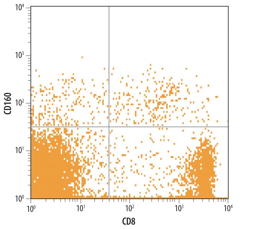
Detection of CD160 in Human Blood Lymphocytes by Flow Cytometry.
Human peripheral blood lymphocytes were stained with Mouse Anti-Human CD160 Monoclonal Antibody (Catalog # MAB6700) followed by Allophycocyanin-conjugated Anti-Mouse IgG Secondary Antibody (Catalog # F0101B) and Mouse Anti-Human CD8a PE-conjugated Monoclonal Antibody (Catalog # FAB1509P). Quadrant markers were set based on control antibody staining (Catalog # MAB0041).Detection of Human CD160 by Functional
Triggering of CD160-GPI is consistent with a positive co-stimulation role. A) Triggering of primary CD4+ T-cells with either plate-bound anti-CD3 (1 μg/ml) and anti-CD28 (0.5 μg/ml) or anti-CD3, anti-CD28 and HVEM-Fc (0.2 μg/ml) in the presence or absence of either anti-HVEM (left panel) or anti-CD160 clone CL1-R2 (right panel). IL-2 was measured in the supernatant by ELISA at 24 h post stimulation. Iso-IgG represents the matched isotype control antibody. P values were determined by two-tailed paired t test (data from three independent healthy donors). B) Left panels: Surface expression of CD160-GPI and CD160-TM on Jurkat-NFAT-Luc cells stably-transfected with CD160 plasmids. Mock-transfected cells (light grey histograms in middle and right panels) were used to set the positive and negative gates for FACS. CD160-TM is weakly detected with CD160-GPI antibodies (BY55 clone). Right panels: Quantitative RT-PCR for CD160-GPI and CD160-TM isoforms in Jurkat cells over-expressing either CD160-GPI or CD160-TM, values are relative to the house-keeping GAPDH gene transcripts (One representative experiment, n = 2). Non-transfected Jurkat (control cells) and HeLa cells were used as additional negative controls for CD160 expression. The left graph represents results with a set of Taqman probes that were not isoform selective and hybrdize both CD160-GPI and CD160-TM to demonstrate similar RNA expression levels, whereas the right graph used a set of probes that were CD160-TM specific to confirm the exclusive expression of the different CD160 isoforms in the two cell lines. C) Simultaneous triggering of TCR and CD160 using magnetic Dynal beads coated with anti-CD3, anti-CD28 and either HVEM-Fc (left panels), CD160 monoclonal antibodies (right panels) or their matched IgGs. Cell activation was monitored by measuring the absolute luciferase counts. Control cells are original Jurkat-NFAT-Luc cells non-transfected with either of the CD160 isoforms. NS: non-stimulated. P values were calculated by non-parametric two-tail t test (Mann–Whitney). Image collected and cropped by CiteAb from the following publication (https://pubmed.ncbi.nlm.nih.gov/25179432), licensed under a CC-BY license. Not internally tested by R&D Systems.Detection of Human CD160 by Functional
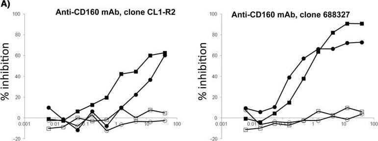
Differential inhibition of HVEM/CD160 binding with benchmark tool antibodies. TRF assay measuring the potency of different antibodies to inhibit the binding of recombinant human HVEM-Fc chimera to CD160+ CHO-K1 cells. A) CD160 monoclonal antibodies inhibit binding of HVEM-Fc to both CD160-GPI and CD160-TM isoforms. B) Polyclonal HVEM (left panel) and monoclonal HVEM (right panel) antibodies both enhance binding of HVEM-Fc to CD160-TM isoform. The polyclonal anti-HVEM inhibits HVEM-Fc binding to CD160-GPI (left panel). Antibody concentrations are plotted on the X axis whereas, the calculated percentage of inhibition of binding is plotted on the Y axis. Matched isotype control antibody for each individual antibody candidate was also used in the assay (empty circles and squares). CTL = control, mAb = monoclonal antibody, pAb = polyclonal antibody. Image collected and cropped by CiteAb from the following publication (https://pubmed.ncbi.nlm.nih.gov/25179432), licensed under a CC-BY license. Not internally tested by R&D Systems.Applications for Human CD160 Antibody
Application
Recommended Usage
CyTOF-ready
Ready to be labeled using established conjugation methods. No BSA or other carrier proteins that could interfere with conjugation.
Flow Cytometry
0.25 µg/106 cells
Sample: Human peripheral blood lymphocytes
Sample: Human peripheral blood lymphocytes
Reviewed Applications
Read 2 reviews rated 4 using MAB6700 in the following applications:
Formulation, Preparation, and Storage
Purification
Protein A or G purified from hybridoma culture supernatant
Reconstitution
Sterile PBS to a final concentration of 0.5 mg/mL. For liquid material, refer to CoA for concentration.
Formulation
Lyophilized from a 0.2 μm filtered solution in PBS with Trehalose. *Small pack size (SP) is supplied either lyophilized or as a 0.2 µm filtered solution in PBS.
Shipping
Lyophilized product is shipped at ambient temperature. Liquid small pack size (-SP) is shipped with polar packs. Upon receipt, store immediately at the temperature recommended below.
Stability & Storage
Use a manual defrost freezer and avoid repeated freeze-thaw cycles.
- 12 months from date of receipt, -20 to -70 °C as supplied.
- 1 month, 2 to 8 °C under sterile conditions after reconstitution.
- 6 months, -20 to -70 °C under sterile conditions after reconstitution.
Background: CD160
V‑type Ig-like domain (aa 27-122). CD160 is found as a soluble, disulfide-linked 80 kDa multimer (likely trimer) that is generated by proteolysis of the GPI-linked form. This 80 kDa form, plus others, are highly resistant to reduction. There is also a 100-110 kDa multimeric transmembrane (TM) form that is associated with activated NK cells. It contains a 55 aa substitution for Gly180-Leu181, and shows a 20 aa TM segment between aa 163-182. The TM form appears to have a splice variant that lacks aa 25-133. Over aa 27-159, human CD160 shares 62% aa identity with mouse CD160.
Alternate Names
BY55, CD160, NK1, NK28
Gene Symbol
CD160
UniProt
Additional CD160 Products
Product Documents for Human CD160 Antibody
Product Specific Notices for Human CD160 Antibody
For research use only
Loading...
Loading...
Loading...
Loading...
Loading...

