Human CD44v6 Antibody
R&D Systems, part of Bio-Techne | Catalog # BBA13

Key Product Details
Validated by
Species Reactivity
Validated:
Cited:
Applications
Validated:
Cited:
Label
Antibody Source
Product Specifications
Immunogen
Specificity
Clonality
Host
Isotype
Scientific Data Images for Human CD44v6 Antibody
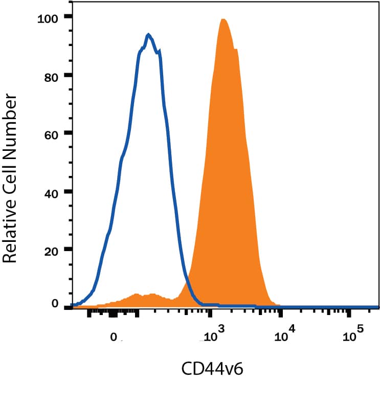
Detection of CD44 in Human Blood Monocytes by Flow Cytometry.
Human peripheral blood monocytes were stained with Mouse Anti-Human CD44 v6 Monoclonal Antibody (Catalog # BBA13, filled histogram) or isotype control antibody (MAB002, open histogram), followed by Allophycocyanin-conjugated Anti-Mouse IgG Secondary Antibody (F0101B).CD44 in A431 Human Cell Line.
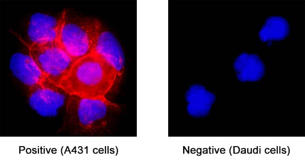
CD44 was detected in immersion fixed A431 human epithelial carcinoma cell line (positive staining) and Daudi human Burkitt's lymphoma cell line (negative staining) using Mouse Anti-Human CD44 v6 Monoclonal Antibody (Catalog # BBA13) at 25 µg/mL for 3 hours at room temperature. Cells were stained using the NorthernLights™ 557-conjugated Anti-Mouse IgG Secondary Antibody (red; NL007) and counterstained with DAPI (blue). Specific staining was localized to cell surface. Staining was performed using our protocol for Fluorescent ICC Staining of Non-adherent Cells.CD44 in Human Colon Adenocarcinoma Tissue.
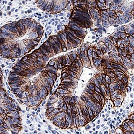
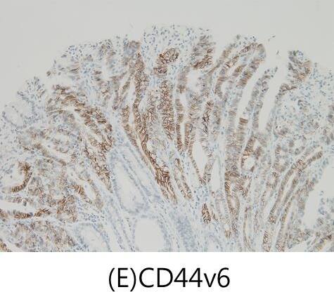
CD44 was detected in immersion fixed paraffin-embedded sections of human colon adenocarcinoma tissue using Mouse Anti-Human CD44 v6 Monoclonal Antibody (Catalog # BBA13) at 1.7 µg/mL for 1 hour at room temperature followed by incubation with the Anti-Mouse IgG VisUCyte™ HRP Polymer Antibody (VC001). Before incubation with the primary antibody, tissue was subjected to heat-induced epitope retrieval using Antigen Retrieval Reagent-Basic (CTS013). Tissue was stained using DAB (brown) and counterstained with hematoxylin (blue). Specific staining was localized to plasma membrane. View our protocol for IHC Staining with VisUCyte HRP Polymer Detection Reagents.Applications for Human CD44v6 Antibody
CyTOF-ready
Flow Cytometry
Sample: Human peripheral blood monocytes
Immunocytochemistry
Sample: Immersion fixed A431 human epithelial carcinoma cell line
Immunohistochemistry
Sample: Immersion fixed paraffin-embedded sections of human colon adenocarcinoma tissue
Immunoprecipitation
Western Blot
Sample: Recombinant Human CD44 v3-10
Reviewed Applications
Read 1 review rated 5 using BBA13 in the following applications:
Formulation, Preparation, and Storage
Purification
Reconstitution
Formulation
Shipping
Stability & Storage
- 12 months from date of receipt, -20 to -70 °C as supplied.
- 1 month, 2 to 8 °C under sterile conditions after reconstitution.
- 6 months, -20 to -70 °C under sterile conditions after reconstitution.
Background: CD44
CD44 is a ubiquitously expressed protein that is the major receptor for hyaluronan and exerts control over cell growth and migration (1‑3). Human CD44 has a 20 amino acid (aa) signal sequence, an extracellular domain (ECD) with a 100 aa hyaluronan‑binding disulfide‑stabilized link region and a 325‑530 aa stem region, a 21 aa transmembrane domain, and a 72 aa cytoplasmic domain. Within the stem, ten variably spliced exons (v1‑10, exons 6‑15) produce multiple protein isoforms (1‑3). The standard or hematopoietic form, CD44H, does not include the variable segments (1‑3). Cancer aggressiveness and T cell activation have been correlated with expression of specific isoforms (1, 3). CD44v6 contains exon 10 and is associated with tumor progression and metastasis in many types of cancer including breast, colon, lung, renal, skin, and ovarian tumors. With variable N‑ and O‑glycosylation and splicing within the stalk, CD44 can range from 80 to 200 kDa (1). Within the N‑terminal invariant portion of the ECD (aa 21‑220), human CD44 shares 76%, 76%, 86%, 83% and 79% identity with corresponding mouse, rat, equine, canine and bovine CD44, respectively. The many reported functions of CD44 fall within three categories (1). First, CD44 binds hyaluronan and other ligands within the extracellular matrix and can function as a “platform” for growth factors and metalloproteinases. Second, CD44 can function as a co‑receptor that modifies activity of receptors including MET and the ERBB family of tyrosine kinases. Third, the CD44 intracellular domain links the plasma membrane to the actin cytoskeleton via the ERM proteins, ezrin, radixin and moesin. CD44 can be synthesized in a soluble form (4) or may be cleaved at multiple sites by either membrane‑type matrix metalloproteinases, or ADAM proteases to produce soluble ectodomains (5, 6). The cellular portion may then undergo gamma secretase‑dependent intramembrane cleavage to form an A beta‑like transmembrane portion and a cytoplasmic signaling portion that affects gene expression (7, 8). These cleavage events are thought to promote metastasis by enhancing tumor cell motility and growth (1, 5).
References
- Ponta, H. et al. (2003) Nat. Rev. Mol. Cell Biol. 4:33.
- Screaton, G.R. et al. (1992) Proc. Natl. Acad. Sci. USA 89:12160.
- Lynch, K.W. (2004) Nat. Rev. Immunol. 4:931.
- Yu, Q. and B.P. Toole (1996) J. Biol. Chem. 271:20603.
- Nagano, O. and H. Saya (2004) Cancer Sci. 95:930.
- Nakamura, H. et al. (2004) Cancer Res. 64:876.
- Murakami, D. et al. (2003) Oncogene 22:1511.
- Lammich, S. et al. (2002) J. Biol. Chem. 277:44754.
- Fox, S.B. et al. (1994) Cancer Res. 54:4539.
Alternate Names
Gene Symbol
Additional CD44 Products
Product Documents for Human CD44v6 Antibody
Product Specific Notices for Human CD44v6 Antibody
For research use only
