Human CD47 Antibody
R&D Systems, part of Bio-Techne | Catalog # AF4670

Key Product Details
Validated by
Species Reactivity
Validated:
Cited:
Applications
Validated:
Cited:
Label
Antibody Source
Product Specifications
Immunogen
Gln19-Pro139
Accession # Q08722
Specificity
Clonality
Host
Isotype
Endotoxin Level
Scientific Data Images for Human CD47 Antibody
Detection of Human CD47 by Western Blot.
fWestern blot shows lysates of U937 human histiocytic lymphoma cell line and human placenta tissue, not heated to minimize aggregation. PVDF membrane was probed with 1 µg/mL of Sheep Anti-Human CD47 Antigen Affinity-purified Polyclonal Antibody (Catalog # AF4670) followed by HRP-conjugated Anti-Sheep IgG Secondary Antibody (Catalog # HAF016). Specific bands were detected for CD47 at approximately 45-70 kDa (as indicated). This experiment was conducted under reducing conditions and using Immunoblot Buffer Group 1.Detection of CD47 in Human Lymphocytes by Flow Cytometry.
Human whole blood lymphocytes were stained with Sheep Anti-Human CD47 Antigen Affinity-purified Polyclonal Antibody (Catalog # AF4670, filled histogram) or control antibody (Catalog # 5-001-A, open histogram), followed by Northern-Lights™ 557-conjugated Anti-Sheep IgG Secondary Antibody (Catalog # NL010).CD47 in Human Placenta.
CD47 was detected in immersion fixed paraffin-embedded sections of human placenta using 5 µg/mL Sheep Anti-Human CD47 Antigen Affinity-purified Polyclonal Antibody (Catalog # AF4670) overnight at 4 °C. Tissue was stained with the Anti-Sheep HRP-DAB Cell & Tissue Staining Kit (brown; Catalog # CTS019) and counterstained with hematoxylin (blue). View our protocol for Chromogenic IHC Staining of Paraffin-embedded Tissue Sections.Applications for Human CD47 Antibody
CyTOF-ready
Flow Cytometry
Sample: Human whole blood lymphocytes
Immunohistochemistry
Sample: Immersion fixed paraffin-embedded sections of human placenta
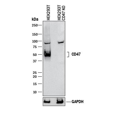
Knockout Validated
Western Blot
Sample: U937 human histiocytic lymphoma cell line and human placenta tissue, not heated to minimize aggregation
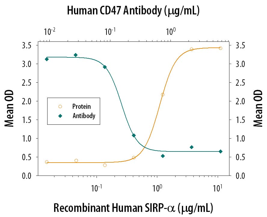
Neutralization
Reviewed Applications
Read 7 reviews rated 4.6 using AF4670 in the following applications:
Formulation, Preparation, and Storage
Purification
Reconstitution
Formulation
Shipping
Stability & Storage
- 12 months from date of receipt, -20 to -70 °C as supplied.
- 1 month, 2 to 8 °C under sterile conditions after reconstitution.
- 6 months, -20 to -70 °C under sterile conditions after reconstitution.
Background: CD47
CD47 (also integrin-associated protein/IAP and OA3) is a variably glycosylated, 40‑60 kDa atypical member of the Ig-Superfamily. It is expressed on almost all cell types, including erythrocytes. CD47 binds to TSP-1 and SIRP alpha, and forms a membrane complex with CD36 and alphav beta3. Mature human CD47 is a 305 amino acid (aa), five-transmembrane glycoprotein. It contains a 123 aa extracellular region (aa 19‑141) that is characterized by the presence of a V-type Ig-like domain (aa 19‑127), and a 34 aa C-terminal cytoplasmic tail that interacts with Gi alpha subunits. Three splice variants occur over aa 293‑323. Over aa 19‑139, human CD47 shares 61%, 71% and 66% aa identity with mouse, porcine and canine CD47, respectively.
Alternate Names
Entrez Gene IDs
Gene Symbol
UniProt
Additional CD47 Products
Product Documents for Human CD47 Antibody
Product Specific Notices for Human CD47 Antibody
For research use only


