Human Complement Component C5aR1 Antibody
R&D Systems, part of Bio-Techne | Catalog # MAB36481


Key Product Details
Species Reactivity
Applications
Label
Antibody Source
Product Specifications
Immunogen
Met1-Val350
Accession # P21730
Specificity
Clonality
Host
Isotype
Scientific Data Images for Human Complement Component C5aR1 Antibody
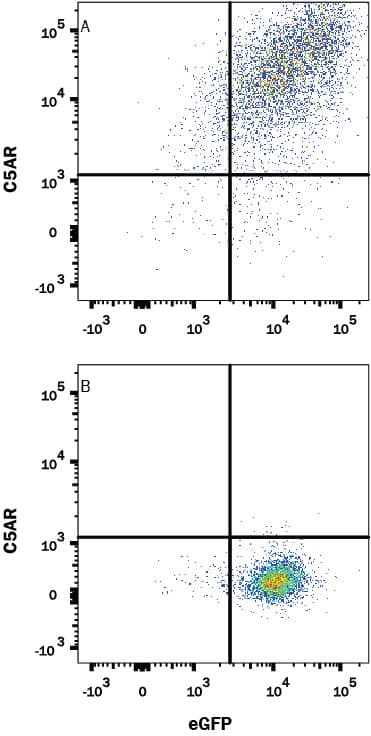
Detection of C5aR in HEK293 Human Cell Line Transfected with Human C5aR and eGFP by Flow Cytometry.
HEK293 human embryonic kidney cell line transfected with either (A) human C5aR or (B) irrelevant protein and eGFP was stained with Mouse Anti-Human C5aR Monoclonal Antibody (Catalog # MAB36481) followed by Allophycocyanin-conjugated Anti-Mouse IgG Secondary Antibody (Catalog # F0101B). Quadrant markers were set based on control antibody staining (Catalog # MAB003). View our protocol for Staining Membrane-associated Proteins.Complement Component C5aR1 in U937 Human Cell Line.
Complement Component C5aR1 was detected in immersion fixed U937 human histiocytic lymphoma cell line (positive) and SH-SY5Y human neuroblastoma cell line (negative control) using Mouse Anti-Human Complement Component C5aR1 Monoclonal Antibody (Catalog # MAB36481) at 8 µg/mL for 3 hours at room temperature. Cells were stained using the NorthernLights™ 557-conjugated Anti-Mouse IgG Secondary Antibody (red; Catalog # NL007) and counterstained with DAPI (blue). Specific staining was localized to cytoplasm. View our protocol for Fluorescent ICC Staining of Cells on Coverslips.Complement Component C5aR1 in Human Spleen.
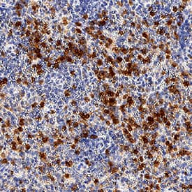
Complement Component C5aR1 was detected in immersion fixed paraffin-embedded sections of human spleen tissue using Mouse Anti-Human Complement Component C5aR1 Monoclonal Antibody (Catalog # MAB36481) at 5 µg/mL for 1 hour at room temperature followed by incubation with the Anti-Mouse IgG VisUCyte™ HRP Polymer Antibody (Catalog # VC001). Before incubation with the primary antibody, tissue was subjected to heat-induced epitope retrieval using Antigen Retrieval Reagent-Basic (Catalog # CTS013). Tissue was stained using DAB (brown) and counterstained with hematoxylin (blue). Specific staining was localized to cytoplasm in splenocytes. View our protocol for IHC Staining with VisUCyte HRP Polymer Detection Reagents.Applications for Human Complement Component C5aR1 Antibody
CyTOF-ready
Flow Cytometry
Sample: HEK293 Human Cell Line Transfected with Human C5aR and eGFP
Immunocytochemistry
Sample: Immersion fixed U937 human histiocytic lymphoma cell line
Immunohistochemistry
Sample: Immersion fixed paraffin-embedded sections of human spleen tissue
Formulation, Preparation, and Storage
Purification
Reconstitution
Formulation
Shipping
Stability & Storage
- 12 months from date of receipt, -20 to -70 °C as supplied.
- 1 month, 2 to 8 °C under sterile conditions after reconstitution.
- 6 months, -20 to -70 °C under sterile conditions after reconstitution.
Background: Complement Component C5aR1
Complement Component C5a R1 (C5aR1), also known as C5a ligand and CD88, is a 7TM protein expressed on myeloid, endothelial, epithelial, and smooth muscle cells. C5aR binds the activated complement anaphylatoxin C5a. In established allergic environments, this triggers neutrophil and eosinophil chemotaxis and the release of proinflammatory mediators. In contrast, C5aR1/C5a interactions are protective during allergen sensitization. Human C5aR1 shares 66% amino acid sequence identity with mouse and rat C5aR.
Long Name
Alternate Names
Gene Symbol
UniProt
Additional Complement Component C5aR1 Products
Product Documents for Human Complement Component C5aR1 Antibody
Product Specific Notices for Human Complement Component C5aR1 Antibody
For research use only

