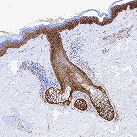
EGFR in Human Skin.
EGFR was detected in immersion fixed frozen sections of human skin using Goat Anti-Human EGFR Antigen Affinity-purified Polyclonal Antibody (Catalog # AF231) at 1 µg/mL for 1 hour at room temperature followed by incubation with the Anti-Goat IgG VisUCyte™ HRP Polymer Antibody (Catalog #
VC004). Tissue was stained using DAB (brown) and counterstained with hematoxylin (blue). Specific staining was localized to plasma membrane. View our protocol for IHC Staining with VisUCyte HRP Polymer Detection Reagents.
Detection of Mouse EGFR by Western Blot
Crizotinib combined with mutant-selective EGFR-TKI overcomes multiple resistances to EGFR-TKI invivo.(A) SCID mice-bearing H1975/Vec- or H1975/HGF- tumors were administered WZ4002 (25 mg/kg) and/or crizotinib (10, 25mg/kg) once daily for 6 to 20 days. Tumor volume was measured using calipers on the indicated days. Mean ± SE tumor volumes are shown for groups of 5 mice. *, P < 0.05 versus control; ✝, P < 0.05 versus WZ4002 by one-way ANOVA. (B) H1975/Vec- or H1975/HGF- tumors were resected from the mice 3 hours after administration of WZ4002 (25mg/kg) and/or crizotinib (10, 25 mg/kg), and the relative levels of proteins in the tumor lysates were determined by western blot analysis. (C) Representative images of H1975/Vec- and H1975/HGF- tumors immunohistochemically stained with antibodies to human Ki-67, and stained with both DAPI (nuclear stain) and TUNEL (FITC). Bar, 200 μm. (D) Quantification of proliferative cells, as determined by the Ki-67-positive proliferation index (percentage of Ki-67-positive cells). Quantification of apoptotic cells, as determined by the TUNEL assay as described in Materials and Methods. Columns, mean of five areas; bars, SD *, P < 0.05 versus of H1975/Vec-tumors; ✝, P < 0.05 versus H1975/HGF-tumors by one-way ANOVA. Image collected and cropped by CiteAb from the following publication (https://dx.plos.org/10.1371/journal.pone.0084700), licensed under a CC-BY license. Not internally tested by R&D Systems.
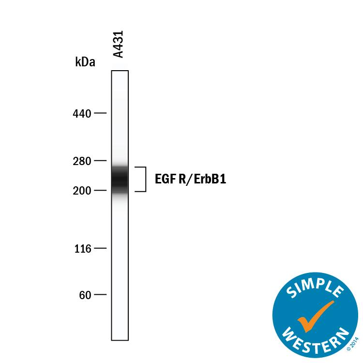
Detection of Human EGFR by Simple WesternTM.
Simple Western lane view shows lysates of A431 human epithelial carcinoma cell line, loaded at 4.2 mg/mL. A specific band was detected for EGFR at approximately 229 kDa (as indicated) using 12.5 µg/mL of Goat Anti-Human EGFR Antigen Affinity-purified Polyclonal Antibody (Catalog # AF231) . This experiment was conducted under reducing conditions and using the 12-230 kDa separation system.
Detection of Human EGFR by Western Blot
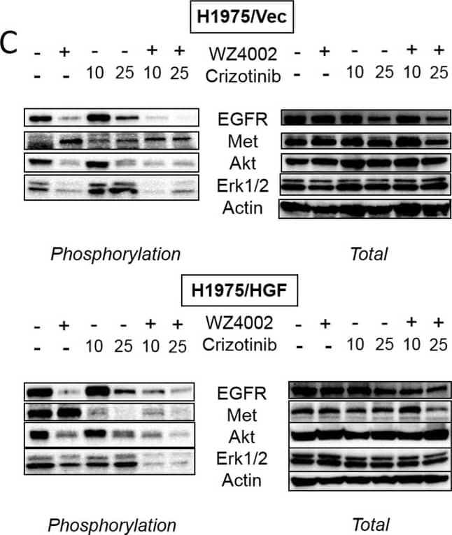
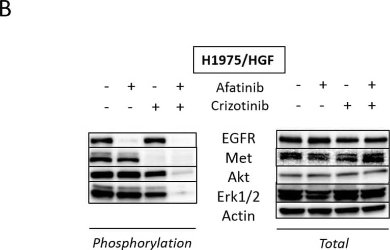
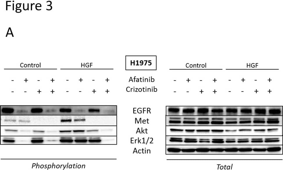
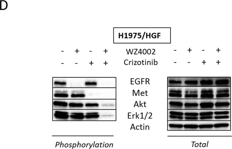
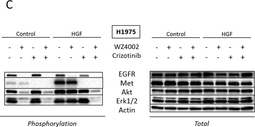
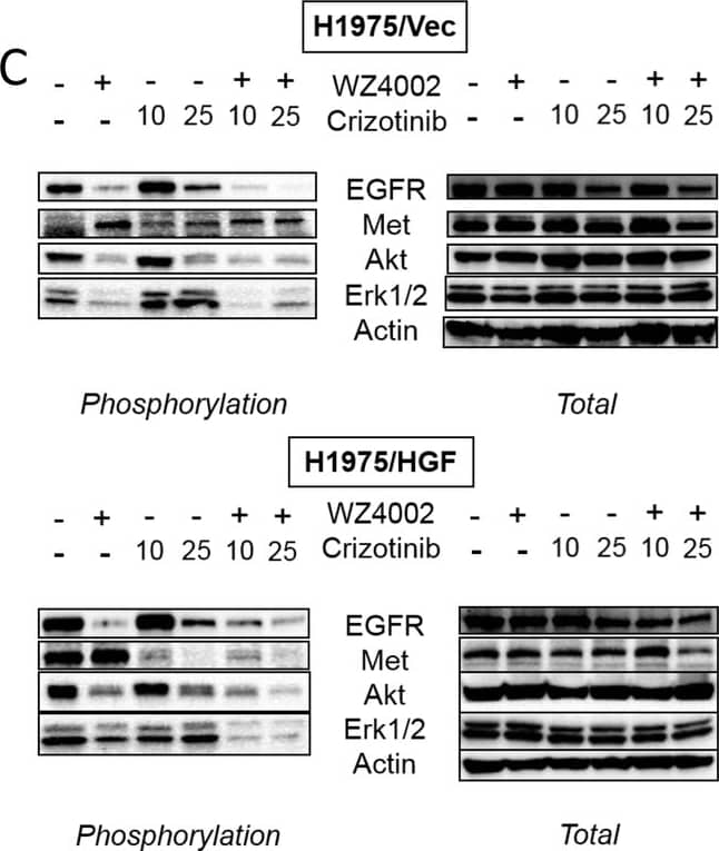
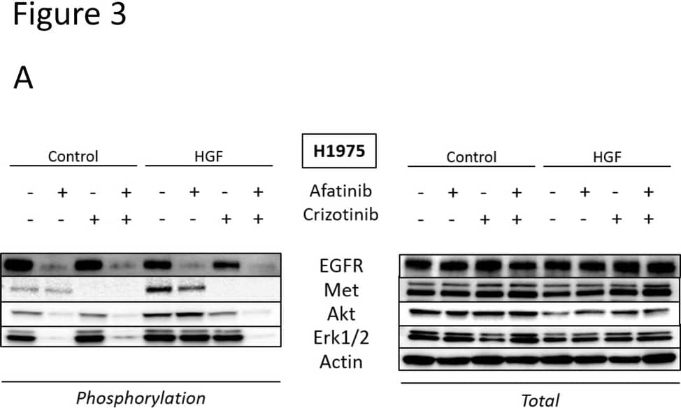
Crizotinib reduces Met phosphorylation and combined treatment with a new generation EGFR-TKI inhibits downstream pathways even in the presence of HGF.H1975 and H1975/HGF cells were incubated with crizotinib (300 nmol/L) and/or afatinib (300 nmol/L) (A, B) or WZ4002 (300 nmol/L) (C, D), for 1 hour. After stimulation with HGF (10 ng/mL) for 10 minutes, the cell lysates were harvested and the phosphorylation of indicated proteins was determined by western blot analysis. Each sample was assayed in triplicate, with each experiment repeated at least 3 times independently. Image collected and cropped by CiteAb from the following publication (https://dx.plos.org/10.1371/journal.pone.0084700), licensed under a CC-BY license. Not internally tested by R&D Systems.
Detection of Mouse EGFR by Western Blot
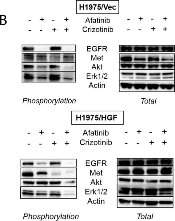
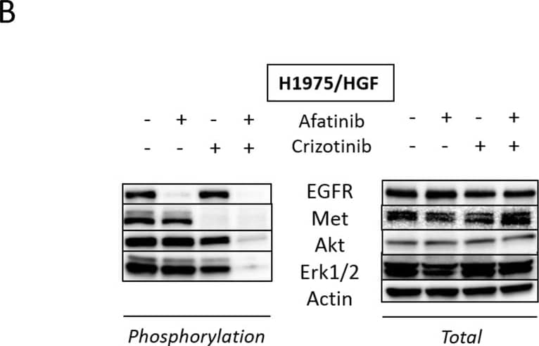
Crizotinib combined with irreversible EGFR-TKI overcomes multiple resistances to EGFR-TKI in vivo.(A) SCID mice-bearing H1975/Vec- or H1975/HGF- tumors were administered afatinib (25 mg/kg) and/or crizotinib (10mg/kg) once daily for 6 to 20 days. Tumor volume was measured using calipers on the indicated days. Mean ± SE tumor volumes are shown for groups of 5 mice. *, P < 0.05 versus control; ✝, P < 0.05 versus afatinib (25 mg/kg) by one-way ANOVA. (B) H1975/Vec- or H1975/HGF- tumors were resected from the mice 3 hours after administration of afatinib (25mg/kg) and/or crizotinib (10 mg/kg), and the relative levels of proteins in the tumor lysates were determined by western blot analysis. (C) Representative images of H1975/Vec- and H1975/HGF tumors immunohistochemically stained with antibodies to human Ki-67, and stained with both DAPI (nuclear stain) and TUNEL (FITC). Bar, 200 μm. (D) Quantification of proliferative cells, as determined by their Ki-67-positive proliferation index (percentage of Ki-67-positive cells). Quantification of apoptotic cells, as determined by the TUNEL assay as described in Materials and Methods. Columns, mean of five areas; bars, SD. *, P < 0.05 versus H1975/Vec-tumors; ✝, P < 0.05 versus control of H1975/HGF-tumors by one-way ANOVA. Image collected and cropped by CiteAb from the following publication (https://dx.plos.org/10.1371/journal.pone.0084700), licensed under a CC-BY license. Not internally tested by R&D Systems.
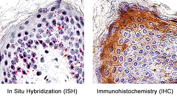
Detection of EGFR in Human Skin.
Formalin-fixed paraffin-embedded tissue sections of human skin were probed for EGFR mRNA (ACD RNAScope Probe, catalog # 310061; Fast Red chromogen, ACD catalog # 322360). Adjacent tissue section was processed for immunohistochemistry using goat anti-human EGFR polyclonal antibody (R&D Systems catalog #
AF231) at 3ug/mL with overnight incubation at 4 degrees Celsius followed by incubation with anti-goat IgG VisUCyte HRP Polymer Antibody (Catalog #
VC004) and DAB chromogen (yellow-brown). Tissue was counterstained with hematoxylin (blue). Specific staining was localized to keratinocytes.
Detection of Human Human EGFR Antibody by Western Blot
Crizotinib reduces Met phosphorylation and combined treatment with a new generation EGFR-TKI inhibits downstream pathways even in the presence of HGF.H1975 and H1975/HGF cells were incubated with crizotinib (300 nmol/L) and/or afatinib (300 nmol/L) (A, B) or WZ4002 (300 nmol/L) (C, D), for 1 hour. After stimulation with HGF (10 ng/mL) for 10 minutes, the cell lysates were harvested and the phosphorylation of indicated proteins was determined by western blot analysis. Each sample was assayed in triplicate, with each experiment repeated at least 3 times independently. Image collected and cropped by CiteAb from the following publication (https://pubmed.ncbi.nlm.nih.gov/24386407), licensed under a CC-BY license. Not internally tested by R&D Systems.
Detection of Mouse Human EGFR Antibody by Western Blot
Crizotinib combined with mutant-selective EGFR-TKI overcomes multiple resistances to EGFR-TKI invivo.(A) SCID mice-bearing H1975/Vec- or H1975/HGF- tumors were administered WZ4002 (25 mg/kg) and/or crizotinib (10, 25mg/kg) once daily for 6 to 20 days. Tumor volume was measured using calipers on the indicated days. Mean ± SE tumor volumes are shown for groups of 5 mice. *, P < 0.05 versus control; ✝, P < 0.05 versus WZ4002 by one-way ANOVA. (B) H1975/Vec- or H1975/HGF- tumors were resected from the mice 3 hours after administration of WZ4002 (25mg/kg) and/or crizotinib (10, 25 mg/kg), and the relative levels of proteins in the tumor lysates were determined by western blot analysis. (C) Representative images of H1975/Vec- and H1975/HGF- tumors immunohistochemically stained with antibodies to human Ki-67, and stained with both DAPI (nuclear stain) and TUNEL (FITC). Bar, 200 μm. (D) Quantification of proliferative cells, as determined by the Ki-67-positive proliferation index (percentage of Ki-67-positive cells). Quantification of apoptotic cells, as determined by the TUNEL assay as described in Materials and Methods. Columns, mean of five areas; bars, SD *, P < 0.05 versus of H1975/Vec-tumors; ✝, P < 0.05 versus H1975/HGF-tumors by one-way ANOVA. Image collected and cropped by CiteAb from the following publication (https://pubmed.ncbi.nlm.nih.gov/24386407), licensed under a CC-BY license. Not internally tested by R&D Systems.
Detection of Human Human EGFR Antibody by Western Blot
Crizotinib reduces Met phosphorylation and combined treatment with a new generation EGFR-TKI inhibits downstream pathways even in the presence of HGF.H1975 and H1975/HGF cells were incubated with crizotinib (300 nmol/L) and/or afatinib (300 nmol/L) (A, B) or WZ4002 (300 nmol/L) (C, D), for 1 hour. After stimulation with HGF (10 ng/mL) for 10 minutes, the cell lysates were harvested and the phosphorylation of indicated proteins was determined by western blot analysis. Each sample was assayed in triplicate, with each experiment repeated at least 3 times independently. Image collected and cropped by CiteAb from the following publication (https://pubmed.ncbi.nlm.nih.gov/24386407), licensed under a CC-BY license. Not internally tested by R&D Systems.


