Human FGFR3 (IIIc) Antibody
R&D Systems, part of Bio-Techne | Catalog # MAB7662
Clone 136312 was used by HLDA to establish CD designation

Key Product Details
Species Reactivity
Validated:
Human
Cited:
Human, Mouse
Applications
Validated:
Flow Cytometry, Immunocytochemistry, Immunohistochemistry, Western Blot
Cited:
ELISA Capture, Immunoprecipitation
Label
Unconjugated
Antibody Source
Monoclonal Mouse IgG1 Clone # 136312
Product Specifications
Immunogen
Pool of NS0-derived recombinant human FGF R3 alpha (IIIb) and Sf21-derived FGF R3 alpha (IIIc)
Specificity
Detects human FGF R3 (IIIc) in direct ELISAs and Western blots. In direct ELISAs, 100% cross‑reactivity with recombinant human (rh) FGF R2 (IIIc), recombinant mouse (rm) FGF R2 (IIIc) and rmFGF R3 (IIIc) is observed. In Western blots (non-reducing conditions only), 50‑100% cross-reactivity with rhFGF R2 (IIIc), rmFGF R2 (IIIc) and rmFGF R3 (IIIc) is observed.
Clonality
Monoclonal
Host
Mouse
Isotype
IgG1
Scientific Data Images for Human FGFR3 (IIIc) Antibody
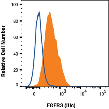
Detection of FGFR3 (IIIc) in HepG2 Human Cell Line by Flow Cytometry.
HepG2 human hepatocellular carcinoma cell line was stained with Mouse Anti-Human FGFR3 (IIIc) Monoclonal Antibody (Catalog # MAB7662, filled histogram) or isotype control antibody (MAB002, open histogram), followed by Allophycocyanin-conjugated Anti-Mouse IgG F(ab')2Secondary Antibody (F0101B). Staining was performed using our Staining Membrane-associated Proteins protocol.FGFR3 in HepG2 Human Cell Line.
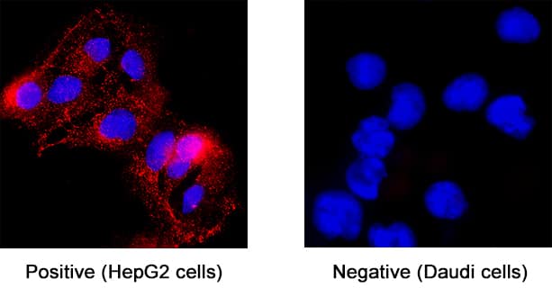
FGFR3 was detected in immersion fixed HepG2 human hepatocellular carcinoma cell line (positive staining) and Daudi human Burkitt's lymphoma cell line (negative staining) using Mouse Anti-Human FGFR3 (IIIc) Monoclonal Antibody (Catalog # MAB7662) at 25 µg/mL for 3 hours at room temperature. Cells were stained using the NorthernLights™ 557-conjugated Anti-Mouse IgG Secondary Antibody (red; NL007) and counterstained with DAPI (blue). Specific staining was localized to cytoplasm. Staining was performed using our protocol for Fluorescent ICC Staining of Non-adherent Cells.FGFR3 in MCF-7 Human Cell Line.
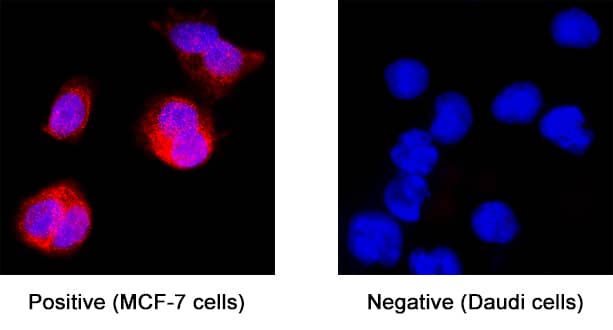
FGFR3 was detected in immersion fixed MCF-7 human breast cancer cell line (positive staining) and Daudi human Burkitt's lymphoma cell line (negative staining) using Mouse Anti-Human FGFR3 (IIIc) Monoclonal Antibody (Catalog # MAB7662) at 25 µg/mL for 3 hours at room temperature. Cells were stained using the NorthernLights™ 557-conjugated Anti-Mouse IgG Secondary Antibody (red; NL007) and counterstained with DAPI (blue). Specific staining was localized to cytoplasm. Staining was performed using our protocol for Fluorescent ICC Staining of Non-adherent Cells.Applications for Human FGFR3 (IIIc) Antibody
Application
Recommended Usage
Flow Cytometry
0.25 µg/106 cells
Sample: HepG2 human hepatocellular carcinoma cell line
Sample: HepG2 human hepatocellular carcinoma cell line
Immunocytochemistry
8-25 µg/mL
Sample: Immersion fixed HepG2 human hepatocellular carcinoma cell line and immersion fixed MCF-7 human breast cancer cell line
Sample: Immersion fixed HepG2 human hepatocellular carcinoma cell line and immersion fixed MCF-7 human breast cancer cell line
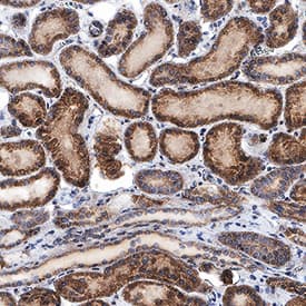
Immunohistochemistry
5-25 µg/mL
Sample: Immersion fixed paraffin-embedded sections of human kidney
Sample: Immersion fixed paraffin-embedded sections of human kidney
Western Blot
1 µg/mL
Sample: Recombinant Human FGF R3 (IIIc) Fc Chimera (Catalog # 766-FR) under non-reducing conditions.
Sample: Recombinant Human FGF R3 (IIIc) Fc Chimera (Catalog # 766-FR) under non-reducing conditions.
Reviewed Applications
Read 1 review rated 3 using MAB7662 in the following applications:
Formulation, Preparation, and Storage
Purification
Protein A or G purified from hybridoma culture supernatant
Reconstitution
Reconstitute at 0.5 mg/mL in sterile PBS. For liquid material, refer to CoA for concentration.
Formulation
Lyophilized from a 0.2 μm filtered solution in PBS with Trehalose. *Small pack size (SP) is supplied either lyophilized or as a 0.2 µm filtered solution in PBS.
Shipping
Lyophilized product is shipped at ambient temperature. Liquid small pack size (-SP) is shipped with polar packs. Upon receipt, store immediately at the temperature recommended below.
Stability & Storage
Use a manual defrost freezer and avoid repeated freeze-thaw cycles.
- 12 months from date of receipt, -20 to -70 °C as supplied.
- 1 month, 2 to 8 °C under sterile conditions after reconstitution.
- 6 months, -20 to -70 °C under sterile conditions after reconstitution.
Background: FGFR3
Long Name
Fibroblast Growth Factor Receptor 3
Alternate Names
CD333, CEK, FGF R3, JTK4
Gene Symbol
FGFR3
Additional FGFR3 Products
Product Documents for Human FGFR3 (IIIc) Antibody
Product Specific Notices for Human FGFR3 (IIIc) Antibody
For research use only
Loading...
Loading...
Loading...
Loading...
Loading...


