Human GDF-9 Antibody
R&D Systems, part of Bio-Techne | Catalog # MAB8266


Key Product Details
Species Reactivity
Applications
Label
Antibody Source
Product Specifications
Immunogen
Met1-Arg454
Accession # O60383
Specificity
Clonality
Host
Isotype
Scientific Data Images for Human GDF-9 Antibody
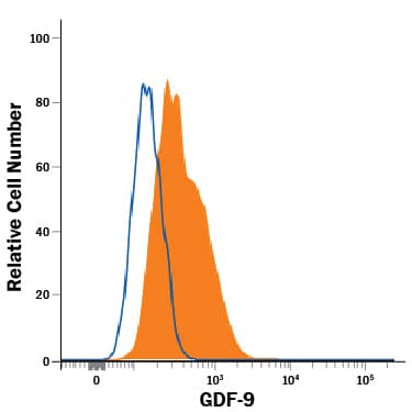
Detection of GDF-9 in OVCAR-3 Human Cell Line by Flow Cytometry.
OVCAR-3 human ovarian carcinoma cell line was stained with Mouse Anti-Human GDF-9 Monoclonal Antibody (Catalog # MAB8266, filled histogram) or isotype control antibody (Catalog # MAB0041, open histogram), followed by Allophycocyanin-conjugated Anti-Mouse IgG Secondary Antibody (Catalog # F0101B). To facilitate intracellular staining, cells were fixed with Flow Cytometry Fixation Buffer (Catalog # FC004) and permeabilized with Flow Cytometry Permeabilization/Wash Buffer I (Catalog # FC005).GDF‑9 in OVCAR‑3 Human Cell Line.
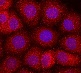
GDF-9 was detected in immersion fixed OVCAR-3 human ovarian carcinoma cell line using Mouse Anti-Human GDF-9 Monoclonal Antibody (Catalog # MAB8266) at 10 µg/mL for 3 hours at room temperature. Cells were stained using the NorthernLights™ 557-conjugated Anti-Mouse IgG Secondary Antibody (red; Catalog # NL007) and counterstained with DAPI (blue). Specific staining was localized to secreted molecule. View our protocol for Fluorescent ICC Staining of Cells on Coverslips.Applications for Human GDF-9 Antibody
CyTOF-ready
Flow Cytometry
Sample: OVCAR‑3 human ovarian carcinoma cell line fixed with Flow Cytometry Fixation Buffer (Catalog # FC004) and permeabilized with Flow Cytometry Permeabilization/Wash Buffer I (Catalog # FC005).
Immunocytochemistry
Sample: Immersion fixed OVCAR‑3 human ovarian carcinoma cell line
Formulation, Preparation, and Storage
Purification
Reconstitution
Formulation
Shipping
Stability & Storage
- 12 months from date of receipt, -20 to -70 °C as supplied.
- 1 month, 2 to 8 °C under sterile conditions after reconstitution.
- 6 months, -20 to -70 °C under sterile conditions after reconstitution.
Background: GDF-9
Growth Differentiation Factor-9 (GDF-9) is an oocyte secreted paracrine factor in the TGF-beta superfamily (1, 2). It is synthesized as a prepropeptide and is subsequently processed by proteases into the mature protein (1, 2). Mature human GDF-9 has a predicted molecular weight of 16 kDa and shares 89.6% and 91.9% amino acid sequence identity with the mouse and rat orthologs, respectively. Despite the high homology, mouse GDF-9 is secreted in an active form, while human GDF-9 is latent. A single mutation Gly391Arg increases the affinity between human GDF-9 and its signaling receptors and make it more active (3). It forms both non-covalent homodimers and heterodimers with BMP-15, which is coordinately expressed with GDF-9 in the oocyte. (2, 4, 5). GDF-9 signals through TGF-beta RI/ALK-5 and BMPR-II, while the GDF-9:BMP-15 heterodimer is believed to signal through BMPR-II, ALK 4/5/7, and BMPR-IB/ALK-6 (5-8). SMAD2 and SMAD3 are phosphorylated following activation of receptor complexes by GDF-9 (5, 6). GDF-9 functions as a paracrine factor in the development of primary follicles in the ovary. It is critical for the growth of granulosa and theca cells and for the differentiation and maturation of the oocyte (5, 9-11). GDF-9 is thought to act synergistically with BMP-15 to control development of the oocyte-cumulus cell complex (4-6). In humans, GDF-9:BMP-15 heterodimers have been shown to be more potent regulators of granulosa cell functions compared to GDF-9 homodimers (6). Aberrant GDF-9 expression and activation is associated with a multitude of common human ovarian disorders including premature ovarian failure and polycystic ovary syndrome (10, 12-14). In breast and bladder cancers, GDF-9 is believed to function as a tumor suppressor because its expression levels are inversely correlated with the aggressiveness of the cancer (15, 16). In prostate cancer, however, GDF-9 may enhance tumor progression by promoting tumor cell growth and epithelial-to-mesenchymal transition (17, 18).
References
- McGrath, S. A. et al. (1995) Mol. Endocrinol. 9:131.
- Aaltonen, J. et al. (1999) J. Clin. Endocrinol. Metab. 84:2744.
- Simpson, C.M. et al. (2012) 153:1301.
- Liao, W.X. et al. (2003) J. Biol. Chem. 278:3713.
- Gilchrist, R.B. et al. (2008) Hum. Reprod. Update 14:159.
- Peng, J. et al. (2013) Proc. Natl. Acad. Sci. USA 110:E776.
- Vitt, U.A. et al. (2002) Biol. Reprod. 67:473.
- Mazerbourg, S. et al. (2004) Mol. Endocrinol. 18:653.
- Hreinsson, J.G. et al. (2002) J. Clin. Endocrinol. Metab. 87:316.
- Otsuka, F. et al. (2011) Mol. Reprod. Dev. 78:9.
- Dong, J. et al. (1996) Nature 383:531.
- Zhao, S.Y. et al. (2010) Fertil. Steril. 94:261.
- Wei, L.N. et al. (2011) Fertil. Steril. 96:464.
- Simpson, C.M. et al. (2014) J. Clin. Endocrinol. Metab. [Epub ahead of print].
- Hanavadi, S. et al. (2007) Ann. Surg. Oncol. 14:2159.
- Du, P. et al. (2012) Int. J. Mol. Med. 29:428.
- Bokobza, S.M. et al. (2010) J. Cell. Physiol. 225:529.
- Bokobza, S.M. et al. (2011) Mol. Cell. Biochem. 349:33.
Long Name
Alternate Names
Gene Symbol
UniProt
Additional GDF-9 Products
Product Documents for Human GDF-9 Antibody
Product Specific Notices for Human GDF-9 Antibody
For research use only
