Human IGFBP-3 Antibody
R&D Systems, part of Bio-Techne | Catalog # MAB11488

Key Product Details
Species Reactivity
Applications
Label
Antibody Source
Product Specifications
Immunogen
Gly28-Lys291
Accession # CAA46087
Specificity
Clonality
Host
Isotype
Scientific Data Images for Human IGFBP-3 Antibody
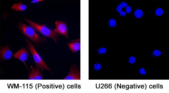
Detection of IGFBP-3 in WM-115 cells (Positive) and U266 cells (Negative).
IGFBP-3 was detected in fixed WM-115 cells (Positive) and absent in U266 human myeloma cell line (Negative) using Mouse Anti-Human IGFBP-3 Monoclonal Antibody (Catalog # mab11488) at 8 µg/ml for 3 hours at room temperature. Cells were stained using the NorthernLights™ 557-conjugated Anti-Mouse IgG Secondary Antibody (red; Catalog # NL007) and counterstained with DAPI (blue). Specific staining was localized to the cytoplasm. View our protocol for Fluorescent ICC Staining of Cells on Coverslips.Detection of IGFBP-3 in Placenta.
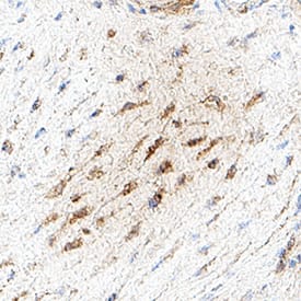
IGFBP-3 was detected in immersion fixed paraffin-embedded sections of placenta using Mouse Anti-Human IGFBP-3 Monoclonal Antibody (Catalog # mab11488) at 5 µg/ml for 1 hour at room temperature followed by incubation with the Anti-Mouse IgG VisUCyte™ HRP Polymer Antibody (Catalog # VC001). Before incubation with the primary antibody, tissue was subjected to heat-induced epitope retrieval using VisUCyte Antigen Retrieval Reagent-Basic (Catalog # VCTS021). Tissue was stained using DAB (brown) and counterstained with hematoxylin (blue). Specific staining was localized to the cytoplasm. View our protocol for Chromogenic IHC Staining of Paraffin-embedded Tissue Sections.Applications for Human IGFBP-3 Antibody
Immunocytochemistry
Sample: Fixed WM-115 cells (Positive) and absent in U266 human myeloma cell line (Negative)
Immunohistochemistry
Sample: Immersion fixed paraffin-embedded sections of placenta
Formulation, Preparation, and Storage
Purification
Reconstitution
Formulation
*Small pack size (-SP) is supplied either lyophilized or as a 0.2 μm filtered solution in PBS. *Small pack size (SP) is supplied either lyophilized or as a 0.2 µm filtered solution in PBS.
Shipping
Stability & Storage
- 12 months from date of receipt, -20 to -70 °C as supplied.
- 1 month, 2 to 8 °C under sterile conditions after reconstitution.
- 6 months, -20 to -70 °C under sterile conditions after reconstitution.
Background: IGFBP-3
The superfamily of insulin-like growth factor (IGF) binding proteins include the six high-affinity IGF binding proteins (IGFBP) and at least four additional low-affinity binding proteins referred to as IGFBP related proteins (IGFBP-rP). All IGFBP superfamily members are cysteine-rich proteins with conserved cysteine residues, which are clustered in the amino- and carboxy-terminal thirds of the molecule. IGFBPs modulate the biological activities of IGF proteins. Some IGFBPs may also have intrinsic bioactivity that is independent of their ability to bind IGF proteins. Post-translational modifications of IGFBPs, including glycosylation, phosphorylation and proteolysis, have been shown to modify the affinities of the binding proteins to IGF.
Human IGFBP-3 cDNA encodes a 291 amino acid (aa) residue precursor protein with a putative 27 aa residue signal peptide that is processed to generate the 264 aa residue mature protein with three potential N-linked and two potential O-linked glycosylation sites. Human IGFBP-3 is expressed in multiple tissues. The highest expression level is found in the non-paranchymal cells of the liver. Expression levels are also higher during extrauterine life and peak during puberty. Human IGFBP-3 is the major IGF binding protein in plasma where it exists in a ternary complex with IGF-I or IGF-II and the acid-labile subunit (ALS).
References
- Jones, J.I. and D.R. Clemmons (1995) Endocrine Rev. 16:3.
- Kelley, K.M. et al. (1996) Int. J. Biochem. Cell Biol. 28:619.
- Spagnoli, A. and R.G. Rosenfeld (1997) Curr. Op. Endocrinology and Diabetes 4:1.
Long Name
Alternate Names
Gene Symbol
UniProt
Additional IGFBP-3 Products
Product Documents for Human IGFBP-3 Antibody
Product Specific Notices for Human IGFBP-3 Antibody
For research use only

