Human IL-1 RII Antibody
R&D Systems, part of Bio-Techne | Catalog # MAB663


Key Product Details
Species Reactivity
Validated:
Cited:
Applications
Validated:
Cited:
Label
Antibody Source
Product Specifications
Immunogen
Phe14-Glu343 (Ser56Gly and Glu297Gly)
Accession # P27930
Specificity
Clonality
Host
Isotype
Endotoxin Level
Scientific Data Images for Human IL-1 RII Antibody
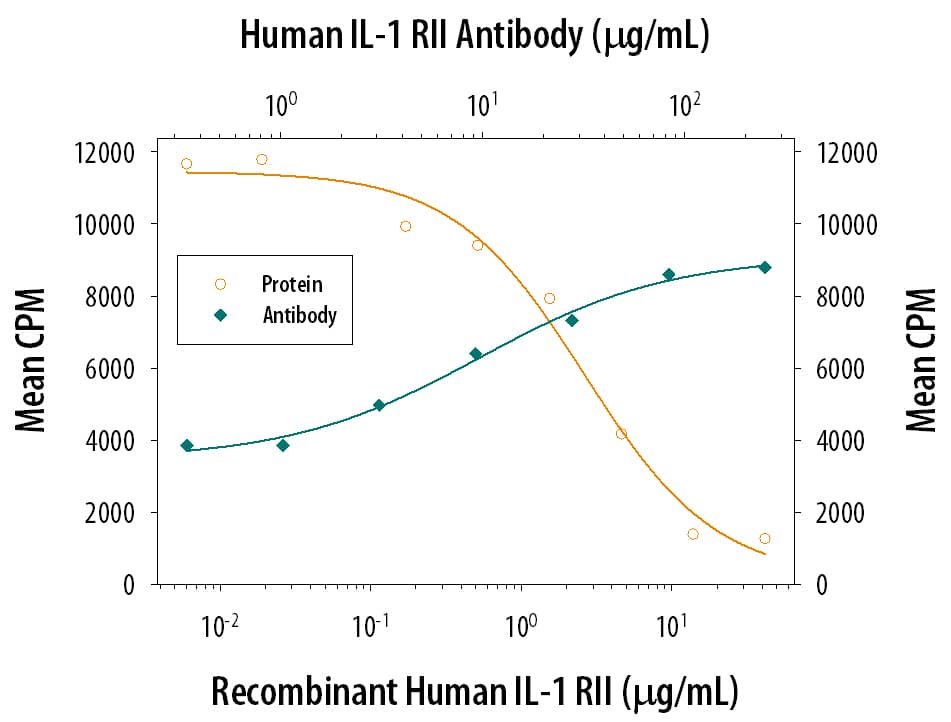
IL-1 RII Inhibition of IL-1 beta/IL-1F2-dependent Cell Proliferation and Neutralization by Human IL-1 RII Antibody.
Recombinant Human IL-1 RII (263-2R) inhibits Recombinant Human IL-1 beta/IL-1F2 (201-LB) induced proliferation in the D10.G4.1 mouse helper T cell line in a dose-dependent manner (orange line). Inhibition of Recombinant Human IL-1 beta/IL-1F2 (50 pg/mL) activity elicited by Recombinant Human IL-1 RII (2 µg/mL) is neutralized (green line) by increasing concentrations of Human IL-1 RII Monoclonal Antibody (Catalog # MAB663). The ND50 is typically 5-20 µg/mL.Detection of IL-1 RII in HDLM-2 cells by Flow Cytometry
HDLM-2 cells were stained with Mouse Anti-Human IL-1 RII Monoclonal Antibody (Catalog # MAB663, filled histogram) or isotype control antibody (Catalog # MAB002, open histogram) followed by Allophycocyanin-conjugated Anti-Mouse IgG Secondary Antibody (Catalog # F0101B). View our protocol for Staining Membrane-associated Proteins.Applications for Human IL-1 RII Antibody
Western Blot
Sample: Recombinant Human IL-1 RII (Catalog # 263-2R)
under non-reducing conditions only
Neutralization
Human IL-1 RII Sandwich Immunoassay
Reviewed Applications
Read 1 review rated 5 using MAB663 in the following applications:
Formulation, Preparation, and Storage
Purification
Reconstitution
Formulation
*Small pack size (-SP) is supplied either lyophilized or as a 0.2 µm filtered solution in PBS.
Shipping
Stability & Storage
- 12 months from date of receipt, -20 to -70 °C as supplied.
- 1 month, 2 to 8 °C under sterile conditions after reconstitution.
- 6 months, -20 to -70 °C under sterile conditions after reconstitution.
Background: IL-1 RII
Two distinct types of receptors that bind the pleiotropic cytokines IL-1 alpha and IL-1 beta have been described. The IL-1 receptor type I is an 80 kDa transmembrane protein that is expressed predominantly by T cells, fibroblasts, and endothelial cells. IL-1 receptor type II is a 68 kDa transmembrane protein found on B lymphocytes, neutrophils, monocytes, large granular leukocytes, and endothelial cells. Both receptors are members of the immunoglobulin superfamily and show approximately 28% sequence similarity in their extracellular domains. The two receptor types do not heterodimerize in a receptor complex. An IL-1 receptor accessory protein that can heterodimerize with the type I receptor in the presence of IL-1 alpha or IL-1 beta, but not IL-1ra, was identified (1). This type I receptor complex appears to mediate all the known IL-1 biological responses. The receptor type II has a short cytoplasmic domain and does not transduce IL-1 signals. In addition to the membrane-bound form of IL-1 RII, a naturally-occurring soluble form of IL-1 RII has been described. It has been suggested that the type II receptor, either as the membrane-bound or as the soluble form, serves as a decoy for IL-1 and inhibits IL-1 action by blocking the binding of IL-1 to the signaling type I receptor complex. Recombinant IL-1 soluble receptor type II is a potent antagonist of IL-1 action.
References
- Greenfeder, S. et al. (1995) J. Biol. Chem. 270:13757.
Long Name
Alternate Names
Gene Symbol
UniProt
Additional IL-1 RII Products
Product Documents for Human IL-1 RII Antibody
Product Specific Notices for Human IL-1 RII Antibody
For research use only
