Human IL-1 RII Fluorescein-conjugated Antibody
R&D Systems, part of Bio-Techne | Catalog # FAB663F


Key Product Details
Validated by
Species Reactivity
Applications
Label
Antibody Source
Product Specifications
Immunogen
Phe14-Glu343 (Ser56Gly and Glu297Gly)
Accession # P27930
Specificity
Clonality
Host
Isotype
Scientific Data Images for Human IL-1 RII Fluorescein-conjugated Antibody
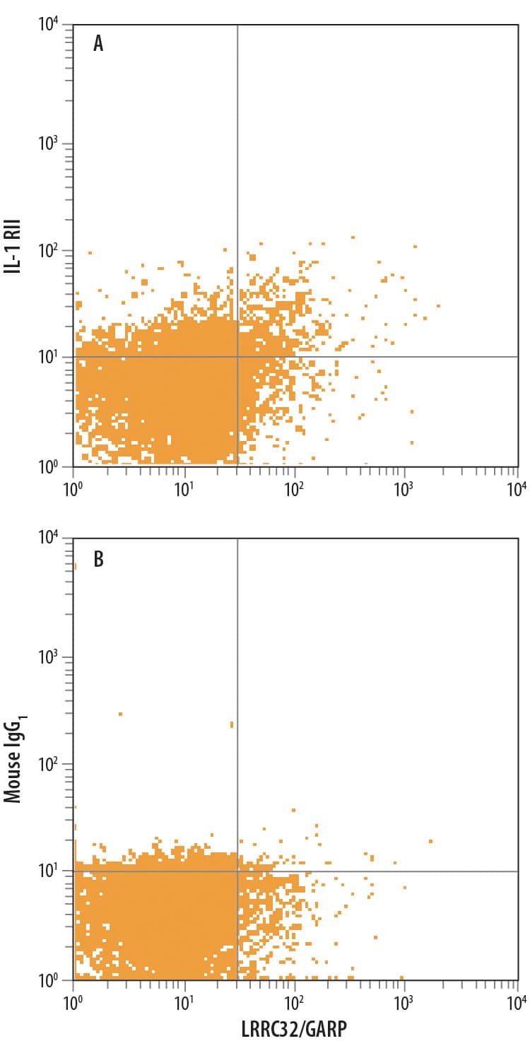
Detection of IL‑1 RII in Human PBMCs stimulated to induce Tregs by Flow Cytometry.
Human peripheral blood mononuclear cells (PBMCs), stimulated to induce Regulatory T Cells (Tregs) and gated on CD4+, were treated with 10 μg/mL Anti-CD3, 5 μg/mL Anti-CD28, 10 ng/mL Recombinant Human TGF-beta 1 (240-B), and 20 ng/mL Recombinant Human IL-2 (202-IL) for 48 hours and stained with Rat Anti-Human LRRC32/GARP APC-conjugated Monoclonal Antibody and either (A) Mouse Anti-Human IL-1 RII Fluorescein-conjugated Monoclonal Antibody (Catalog # FAB663F) or (B) Mouse IgG1Fluorescein Isotype Control (IC002F). View our protocol for Staining Membrane-associated Proteins.Detection of IL-1 RII in HDLM-2 cells by Flow Cytometry
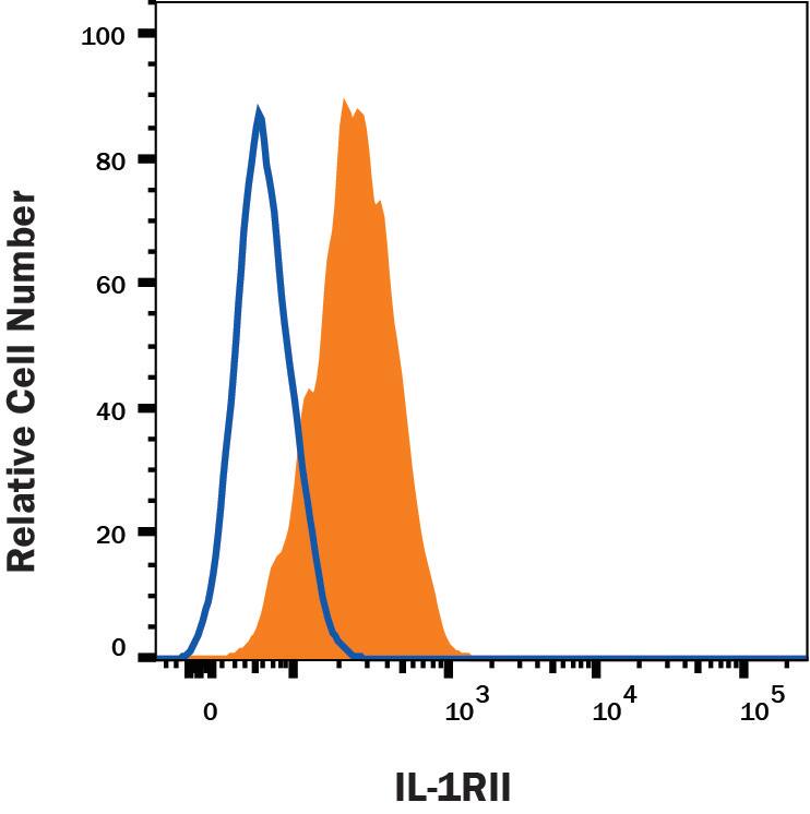
HDLM-2 cells were stained with Mouse Anti-Human IL-1 RII Fluorescein-conjugated Monoclonal Antibody (Catalog # FAB663F, filled histogram) or isotype control antibody (Catalog # IC002F, open histogram). View our protocol for Staining Membrane-associated Proteins.Applications for Human IL-1 RII Fluorescein-conjugated Antibody
Flow Cytometry
Sample: - Human peripheral blood mononuclear cells (PBMCs), stimulated to induce Regulatory T Cells (Tregs) and gated on CD4+, were treated with Anti-CD3, Anti-CD28, Recombinant Human TGF‑ beta1 (Catalog # 240-B), and Recombinant Human IL‑2 (Catalog # 202-IL)
- HDLM-2 cells
Reviewed Applications
Read 1 review rated 5 using FAB663F in the following applications:
Formulation, Preparation, and Storage
Purification
Formulation
Shipping
Stability & Storage
- 12 months from date of receipt, 2 to 8 °C as supplied.
Background: IL-1 RII
Two distinct types of receptors that bind the pleiotropic cytokines IL-1 alpha and IL-1 beta have been described. The IL-1 receptor type I is an 80 kDa transmembrane protein that is expressed predominantly by T cells, fibroblasts, and endothelial cells. IL-1 receptor type II is a 68 kDa transmembrane protein found on B lymphocytes, neutrophils, monocytes, large granular leukocytes, and endothelial cells. Both receptors are members of the immunoglobulin superfamily and show approximately 28% sequence similarity in their extracellular domains. The two receptor types do not heterodimerize in a receptor complex. An IL-1 receptor accessory protein that can heterodimerize with the type I receptor in the presence of IL-1 alpha or IL-1 beta, but not IL-1ra, was identified (1). This type I receptor complex appears to mediate all the known IL-1 biological responses. The receptor type II has a short cytoplasmic domain and does not transduce IL-1 signals. In addition to the membrane-bound form of IL-1 RII, a naturally-occurring soluble form of IL-1 RII has been described. It has been suggested that the type II receptor, either as the membrane-bound or as the soluble form, serves as a decoy for IL-1 and inhibits IL-1 action by blocking the binding of IL-1 to the signaling type I receptor complex. Recombinant IL-1 soluble receptor type II is a potent antagonist of IL-1 action.
References
- Greenfeder, S. et al. (1995) J. Biol. Chem. 270:13757.
Long Name
Alternate Names
Gene Symbol
UniProt
Additional IL-1 RII Products
Product Documents for Human IL-1 RII Fluorescein-conjugated Antibody
Product Specific Notices for Human IL-1 RII Fluorescein-conjugated Antibody
For research use only
