Human IL-10 Antibody
R&D Systems, part of Bio-Techne | Catalog # AF-217-NA


Key Product Details
Species Reactivity
Validated:
Cited:
Applications
Validated:
Cited:
Label
Antibody Source
Product Specifications
Immunogen
Ser19-Asn178
Accession # P22301
Specificity
Clonality
Host
Isotype
Endotoxin Level
Scientific Data Images for Human IL-10 Antibody
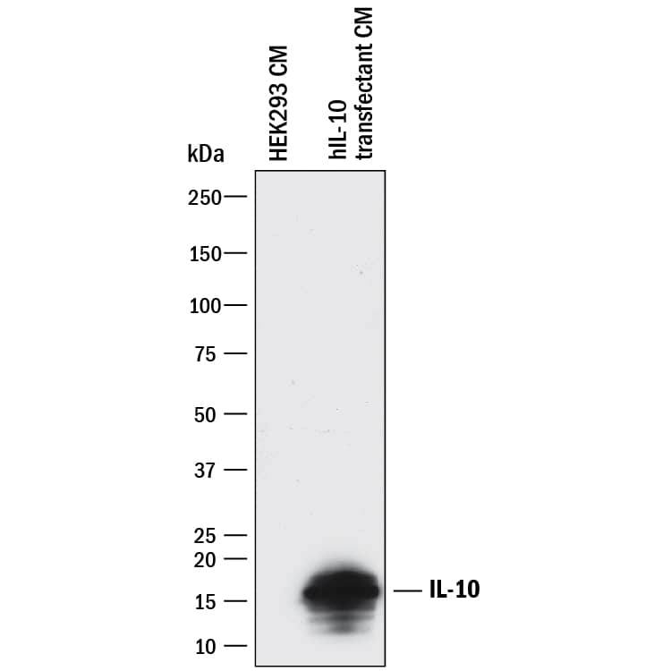
Detection of Human IL‑10 by Western Blot.
Western blot shows conditioned media of HEK293 human embryonic kidney cell line either mock transfected or transfected with human IL-10. PVDF membrane was probed with 1 µg/mL of Goat Anti-Human IL-10 Antigen Affinity-purified Polyclonal Antibody (Catalog # AF-217-NA) followed by HRP-conjugated Anti-Goat IgG Secondary Antibody (Catalog # HAF017). A specific band was detected for IL-10 at approximately 16 kDa (as indicated). This experiment was conducted under reducing conditions and using Immunoblot Buffer Group 1.IL‑10 in Human Tonsil.
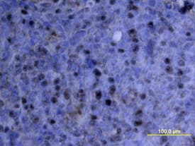
IL-10 was detected in immersion fixed paraffin-embedded sections of human tonsil using Goat Anti-Human IL-10 Antigen Affinity-purified Polyclonal Antibody (Catalog # AF-217-NA) at 15 µg/mL overnight at 4 °C. Tissue was stained using the Anti-Goat HRP-DAB Cell & Tissue Staining Kit (brown; Catalog # CTS008) and counterstained with hematoxylin (blue). View our protocol for Chromogenic IHC Staining of Paraffin-embedded Tissue Sections.IL‑10 in Human Tonsil.
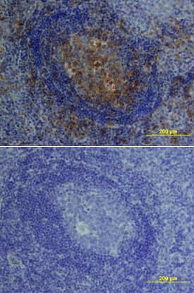
IL-10 was detected in immersion fixed paraffin-embedded sections of human tonsil using Goat Anti-Human IL-10 Antigen Affinity-purified Polyclonal Antibody (Catalog # AF-217-NA) at 15 µg/mL overnight at 4 °C. Tissue was stained using the Anti-Goat HRP-DAB Cell & Tissue Staining Kit (brown; Catalog # CTS008) and counterstained with hematoxylin (blue). Lower panel shows a lack of labeling if primary antibodies are omitted and tissue is stained only with secondary antibody followed by incubation with detection reagents. View our protocol for Chromogenic IHC Staining of Paraffin-embedded Tissue Sections.Applications for Human IL-10 Antibody
Immunohistochemistry
Sample: Immersion fixed paraffin-embedded sections of human tonsil
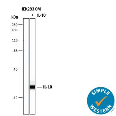
Simple Western
Sample: HEK293 human embryonic kidney cell line transfected with human IL-10
Western Blot
Sample: HEK293 human embryonic kidney cell line transfected with human IL-10
Neutralization
Reviewed Applications
Read 2 reviews rated 4 using AF-217-NA in the following applications:
Formulation, Preparation, and Storage
Purification
Reconstitution
Formulation
Shipping
Stability & Storage
- 12 months from date of receipt, -20 to -70 °C as supplied.
- 1 month, 2 to 8 °C under sterile conditions after reconstitution.
- 6 months, -20 to -70 °C under sterile conditions after reconstitution.
Background: IL-10
Interleukin 10, also known as cytokine synthesis inhibitory factor (CSIF), is the charter member of the IL-10 family of alpha-helical cytokines that also includes IL-19, IL‑20, IL-22, IL-24, and IL-26/AK155 (1, 2). IL-10 is secreted by many activated hematopoietic cell types as well as hepatic stellate cells, keratinocytes, and placental cytotrophoblasts (2‑5). Mature human IL-10 shares 72%‑86% amino acid sequence identity with bovine, canine, equine, feline, mouse, ovine, porcine, and rat IL-10. Whereas human IL-10 is active on mouse cells, mouse IL-10 does not act on human cells (6, 7). IL-10 is a 178 amino acid molecule that contains two intrachain disulfide bridges and is expressed as a 36 kDa noncovalently associated homodimer (6, 8, 9). The IL-10 dimer binds to two IL-10 R alpha/IL-10 R1 chains, resulting in recruitment of two IL-10 R beta/IL-10 R2 chains and activation of a signaling cascade involving JAK1, TYK2, and STAT3 (10). IL-10 R beta does not bind IL-10 by itself but is required for signal transduction (1). IL-10 R beta also associates with IL‑20 R alpha, IL-22 R alpha, or IL-28 R alpha to form the receptor complexes for IL-22, IL-26, IL-28, and IL‑29 (11‑13). IL-10 is a critical molecule in the control of viral infections and allergic and autoimmune inflammation (14‑16). It promotes phagocytic uptake and Th2 responses but suppresses antigen presentation and Th1 proinflammatory responses (2).
References
- Pestka, S. et al. (2004) Annu. Rev. Immunol. 22:929.
- O’Garra, A. and P. Vieira (2007) Nat. Rev. Immunol. 7:425.
- Mathurin, P. et al. (2002) Am. J. Physiol. Gastrointest. Liver Physiol. 282:G981.
- Grewe, M. et al. (1995) J. Invest. Dermatol. 104:3.
- Szony, B.J. et al. (1999) Mol. Hum. Reprod. 5:1059.
- Vieira, P. et al. (1991) Proc. Natl. Acad. Sci. 88:1172.
- Hsu, D.-H. et al. (1990) Science 250:830.
- Windsor, W.T. et al. (1993) Biochemistry 32:8807.
- Syto, R. et al. (1998) Biochemistry 37:16943.
- Kotenko, S.V. et al. (1997) EMBO J. 16:5894.
- Kotenko, S.V. et al. (2000) J. Biol. Chem. 276:2725.
- Hor, S. et al. (2004) J. Biol. Chem. 279:33343.
- Sheppard, P. et al. (2003) Nat. Immunol. 4:63.
- Fitzgerald, D.C. et al. (2007) Nat. Immunol. 8:1372.
- Wu, K. et al. (2007) Cell. Mol. Immunol. 4:269.
- Blackburn, S.D. and E.J. Wherry (2007) Trends Microbiol. 15:143.
Long Name
Alternate Names
Entrez Gene IDs
Gene Symbol
UniProt
Additional IL-10 Products
Product Documents for Human IL-10 Antibody
Product Specific Notices for Human IL-10 Antibody
For research use only


