Human IL-2 Antibody
R&D Systems, part of Bio-Techne | Catalog # MAB202R
Recombinant Monoclonal Antibody.


Conjugate
Catalog #
Key Product Details
Validated by
Biological Validation
Species Reactivity
Human
Applications
Immunocytochemistry, Intracellular Staining by Flow Cytometry, Neutralization
Label
Unconjugated
Antibody Source
Monoclonal Mouse IgG1 Clone # 5334R
Product Specifications
Immunogen
E. coli-derived recombinant human IL-2
Ala21-Thr153
Accession # NP_000577
Ala21-Thr153
Accession # NP_000577
Specificity
Detects human IL-2 in direct ELISAs.
Clonality
Monoclonal
Host
Mouse
Isotype
IgG1
Scientific Data Images for Human IL-2 Antibody
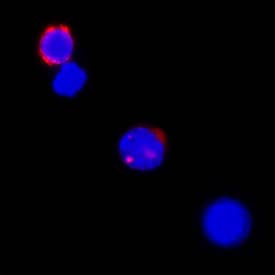
IL-2 in Human PBMCs.
IL-2 was detected in immersion fixed human peripheral blood mononuclear cells (PBMCs) treated with calcium ionomycin and PMA using Mouse Anti-Human IL-2 Monoclonal Antibody (Catalog # MAB202R) at 25 µg/mL for 3 hours at room temperature. Cells were stained using the NorthernLights™ 557-conjugated Anti-Mouse IgG Secondary Antibody (red; Catalog # NL007) and counterstained with DAPI (blue). Specific staining was localized to cytoplasm. View our protocol for Fluorescent ICC Staining of Non-adherent Cells.Cell Proliferation Induced by IL-2 and Neutralization by Human IL-2 Antibody.
Recombinant Human IL-2 (Catalog # 202-IL) stimulates proliferation in the CTLL-2 mouse cytotoxic T cell line in a dose-dependent manner (orange line). Proliferation elicited by Recombinant Human IL-2 (2 ng/mL) is neutralized (green line) by increasing concentrations of Mouse Anti-Human IL-2 Monoclonal Antibody (Catalog # MAB202). The ND50 is typically 0.015-0.03 µg/mL.Detection of IL-2 in Human PBMCs by Flow Cytometry.
Human peripheral blood mononuclear cells (PBMCs) (A) treated with Cell Activation Cocktail 500x (5476) for 5 hours or (B) resting were stained with Mouse Anti-Human IL-2 Monoclonal Antibody (Catalog # MAB202R) followed by Goat anti-Mouse IgG APC-conjugated Secondary Antibody (F0101B) and Mouse anti-Human CD3 PE-conjugated Monoclonal Antibody (FAB100P). Quadrant markers were set based on Mouse IgG1 isotype control antibody (MAB002) . To facilitate intracellular staining, cells were fixed and permeabilized using FlowX FoxP3/Transcription Factor Fixation & Perm Buffer Kit (FC012). Staining was performed using our Staining Intracellular Molecules protocol.Applications for Human IL-2 Antibody
Application
Recommended Usage
Immunocytochemistry
5-25 µg/mL
Sample: Immersion fixed human peripheral blood mononuclear cells (PBMCs) treated with calcium ionomycin and PMA
Sample: Immersion fixed human peripheral blood mononuclear cells (PBMCs) treated with calcium ionomycin and PMA
Intracellular Staining by Flow Cytometry
0.25 µg/106 cells
Sample: Human peripheral blood mononuclear cells (PBMCs) treated with Cell Activation Cocktail 500x (Catalog # 5476) for 5 hours, fixed and permeabilized using FlowX FoxP3/Transcription Factor Fixation & Perm Buffer Kit (Catalog # FC012)
Sample: Human peripheral blood mononuclear cells (PBMCs) treated with Cell Activation Cocktail 500x (Catalog # 5476) for 5 hours, fixed and permeabilized using FlowX FoxP3/Transcription Factor Fixation & Perm Buffer Kit (Catalog # FC012)
Neutralization
Measured by its ability to neutralize IL‑2-induced proliferation in the CTLL‑2 mouse cytotoxic T cell line. Gearing, A.J.H. and C.B. Bird (1987) in Lymphokines and Interferons, A Practical Approach. Clemens, M.J. et al. (eds): IRL Press. 276. The Neutralization Dose (ND50) is typically 0.015-0.03 µg/mL in the presence of 2 ng/mL Recombinant Human IL‑2.
Formulation, Preparation, and Storage
Purification
Protein A or G purified from cell culture supernatant
Reconstitution
Reconstitute at 0.5 mg/mL in sterile PBS. For liquid material, refer to CoA for concentration.
Formulation
Lyophilized from a 0.2 μm filtered solution in PBS with Trehalose. *Small pack size (SP) is supplied either lyophilized or as a 0.2 µm filtered solution in PBS.
Shipping
Lyophilized product is shipped at ambient temperature. Liquid small pack size (-SP) is shipped with polar packs. Upon receipt, store immediately at the temperature recommended below.
Stability & Storage
Use a manual defrost freezer and avoid repeated freeze-thaw cycles.
- 12 months from date of receipt, -20 to -70 °C as supplied.
- 1 month, 2 to 8 °C under sterile conditions after reconstitution.
- 6 months, -20 to -70 °C under sterile conditions after reconstitution.
Background: IL-2
Interleukin-2 (IL-2) is a cytokine that stimulates the growth and differentiation of B cells, T cells, NK cells, and monocyte/macrophages. It functions through the heterotrimeric IL-2 receptor comprising alpha, beta, and gamma chains.
Long Name
Interleukin 2
Alternate Names
Aldesleukin, IL2, Proleukin, TCGF
Entrez Gene IDs
Gene Symbol
IL2
UniProt
Additional IL-2 Products
Product Documents for Human IL-2 Antibody
Product Specific Notices for Human IL-2 Antibody
For research use only
Loading...
Loading...
Loading...
Loading...
Loading...

