Human/Mouse SSEA-1 Antibody
R&D Systems, part of Bio-Techne | Catalog # MAB2155


Conjugate
Catalog #
Key Product Details
Species Reactivity
Validated:
Human, Mouse
Cited:
Human, Mouse, Canine, Chicken, Equine
Applications
Validated:
CyTOF-reported, Flow Cytometry, Immunocytochemistry, Immunoprecipitation
Cited:
Flow Cytometry, Immunocytochemistry, Immunocytochemistry/ Immunofluorescence, Immunohistochemistry, Immunohistochemistry-Paraffin, Western Blot
Label
Unconjugated
Antibody Source
Monoclonal Mouse IgM Clone # MC-480
Product Specifications
Immunogen
F9 mouse teratocarcinoma stem cells
Specificity
Detects human and mouse SSEA-1.
Clonality
Monoclonal
Host
Mouse
Isotype
IgM
Scientific Data Images for Human/Mouse SSEA-1 Antibody
SSEA-1 in D3 Mouse Cell Line.
SSEA-1 was detected in immersion fixed D3 mouse embryonic stem cell line using 10 µg/mL Human/Mouse SSEA-1 Monoclonal Antibody (Catalog # MAB2155) for 3 hours at room temperature. Cells were stained (red) and counterstained with DAPI (blue). View our protocol for Fluorescent ICC Staining of Cells on Coverslips.Detection of SSEA-1 in Whole blood granulocytes by Flow Cytometry.
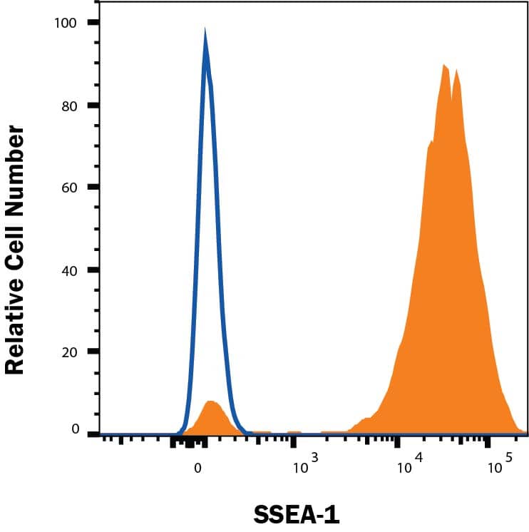
Whole blood granulocytes cells were stained with Mouse Anti-Human/Mouse SSEA-1 Monoclonal Antibody (Catalog # MAB2155, filled histogram) or isotype control antibody (open histogram), followed by Allophycocyanin-conjugated Anti-Mouse IgM Secondary Antibody (Catalog # F0117). View our protocol for Staining Membrane-associated Proteins.Detection of Mouse SSEA-1 by Immunocytochemistry/Immunofluorescence
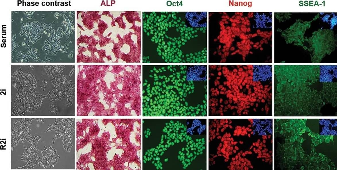
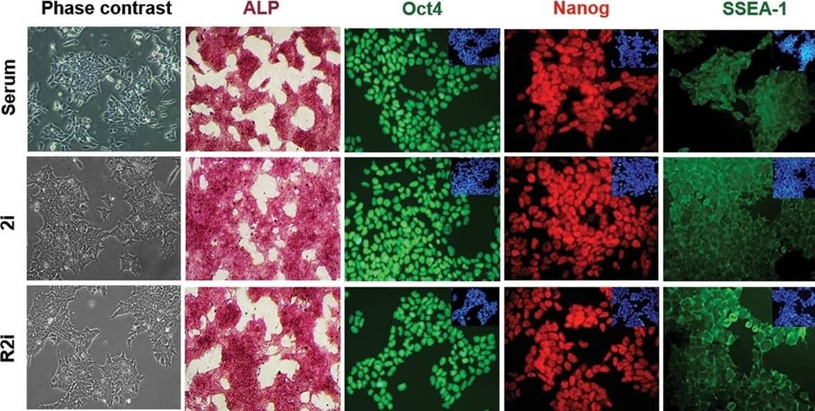
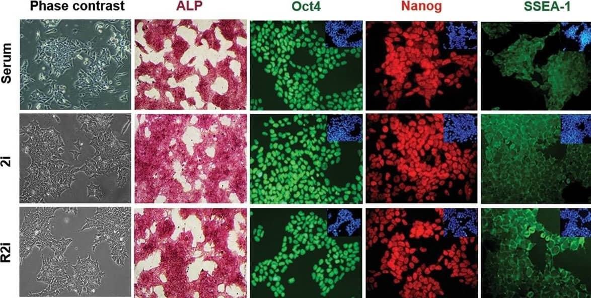
Characteristics of mouse embryonic stem cells (mESCs) cultivated in 2i, R2i, and serum. Alkaline phosphatase (ALP) staining (scale bar: 100 µm) and immunofluorescence (IF) labeling for Oct4, SSEA-1, and Nanog counterstained for DAPI are shown (scale bar: 50 µm). Image collected and cropped by CiteAb from the following publication (https://pubmed.ncbi.nlm.nih.gov/29845793), licensed under a CC-BY license. Not internally tested by R&D Systems.Applications for Human/Mouse SSEA-1 Antibody
Application
Recommended Usage
CyTOF-reported
Lujan, E. et al. (2015) Nature 521: 352. Ready to be labeled using established conjugation methods. No BSA or other carrier proteins that could interfere with conjugation.
Flow Cytometry
0.25 µg/106 cells
Sample: Whole blood granulocytes
Sample: Whole blood granulocytes
Immunocytochemistry
8-25 µg/mL
Sample: Immersion fixed D3 mouse embryonic stem cell line
Sample: Immersion fixed D3 mouse embryonic stem cell line
Immunoprecipitation
Ballou, B. et al. (1979) Science 206:844.
Reviewed Applications
Read 2 reviews rated 4.5 using MAB2155 in the following applications:
Formulation, Preparation, and Storage
Purification
IgM-specific Affinity-purified from hybridoma culture supernatant
Reconstitution
Reconstitute at 0.5 mg/mL in sterile PBS. For liquid material, refer to CoA for concentration.
Formulation
Lyophilized from a 0.2 μm filtered solution in PBS with Trehalose. *Small pack size (SP) is supplied either lyophilized or as a 0.2 µm filtered solution in PBS.
Shipping
Lyophilized product is shipped at ambient temperature. Liquid small pack size (-SP) is shipped with polar packs. Upon receipt, store immediately at the temperature recommended below.
Stability & Storage
Use a manual defrost freezer and avoid repeated freeze-thaw cycles.
- 12 months from date of receipt, -20 to -70 °C as supplied.
- 1 month, 2 to 8 °C under sterile conditions after reconstitution.
- 6 months, -20 to -70 °C under sterile conditions after reconstitution.
Background: SSEA-1
References
- Solter, D. and Knowles, B.B. (1978) Proc. Natl. Acad. Sci. USA 75:5565.
- Fox, N. et al. (1983) Cancer Res. 43:669.
Long Name
Stage-specific Embryonic Antigen-1
Alternate Names
SSEA1
Additional SSEA-1 Products
Product Documents for Human/Mouse SSEA-1 Antibody
Product Specific Notices for Human/Mouse SSEA-1 Antibody
For research use only
Loading...
Loading...
Loading...
Loading...
Loading...


