Human/Mouse SSEA-4 Antibody
R&D Systems, part of Bio-Techne | Catalog # MAB1435


Key Product Details
Species Reactivity
Validated:
Cited:
Applications
Validated:
Cited:
Label
Antibody Source
Product Specifications
Immunogen
Specificity
Clonality
Host
Isotype
Scientific Data Images for Human/Mouse SSEA-4 Antibody
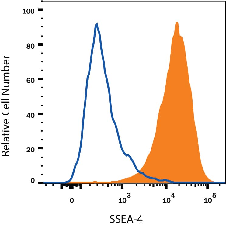
Detection of SSEA‑4 in NTera‑2 Human Cell Line by Flow Cytometry.
NTera-2 human testicular embryonic carcinoma cell line was stained with Mouse Anti-Human/Mouse SSEA-4 Monoclonal Antibody (Catalog # MAB1435, filled histogram) or isotype control antibody (Catalog # MAB007, open histogram), followed by Phycoerythrin-conjugated Anti-Mouse IgG Secondary Antibody (Catalog # F0102B).SSEA-4 in BG01V Human Stem Cells.
SSEA-4 was detected in immersion fixed BG01V human embryonic stem cells cultured on irradiated mouse embryonic fibroblasts (Catalog # PSC001) using 10 µg/mL Mouse Anti-Human/Mouse SSEA-4 Monoclonal Antibody (Catalog # MAB1435) for 3 hours at room temperature. Cells were stained with the NorthernLights™ 557-conjugated Anti-Mouse IgG Secondary Antibody (red; Catalog # NL007) and counterstained with DAPI (blue). View our protocol for Fluorescent ICC Staining of Cells on Coverslips.Detection of Canine SSEA-4 by Flow Cytometry
Flow cytometry. Comparison of cell surface proteins CD29, CD44, CD90, CD34, CD45, SSEA-1, SSEA-3, SSEA-4, TRA-1-60, and TRA-1-81 on primary cultures of BM-MSCs (A, C) and AT-MSCs (B, D). Solid histograms show nonspecific staining and open histograms show specific staining for the indicated marker. Three different donor MSC populations from each tissue type were analyzed and representative samples are shown. Image collected and cropped by CiteAb from the following publication (https://bmcvetres.biomedcentral.com/articles/10.1186/1746-6148-8-150), licensed under a CC-BY license. Not internally tested by R&D Systems.Applications for Human/Mouse SSEA-4 Antibody
Flow Cytometry
Sample: NTera-2 human testicular embryonic carcinoma cell line
Immunocytochemistry
Sample: Immersion fixed BG01V human embryonic stem cells cultured on irradiated mouse embryonic fibroblasts (Catalog # PSC001)
Reviewed Applications
Read 9 reviews rated 4.2 using MAB1435 in the following applications:
Formulation, Preparation, and Storage
Purification
Reconstitution
Formulation
Shipping
Stability & Storage
- 12 months from date of receipt, -20 to -70 °C as supplied.
- 1 month, 2 to 8 °C under sterile conditions after reconstitution.
- 6 months, -20 to -70 °C under sterile conditions after reconstitution.
Background: SSEA-4
SSEA-4 is expressed on the surface of human embryonal carcinoma (EC) cells (the pluripotent stem cells of teratocarcinomas), human embryonic germ cells (EG), and human embryonic stem cells (ES). Expression of SSEA-4 is down-regulated following differentiation of human EC cells. In contrast, the differentiation of murine EC and ES cells may be accompanied by an increase in SSEA-4 expression (1-4).
References
- Shevinsky, L.H. et al. (1982) Cell 30:697.
- Kannagi, R. et al. (1983) EMBO J. 2:2355.
- Thomson, J.A. and J.S. Odorico (2000) Trends Biotechnol. 18:53.
- Draper, J.S. et al. (2002) J. Anat. 200:249.
Long Name
Alternate Names
Additional SSEA-4 Products
Product Documents for Human/Mouse SSEA-4 Antibody
Product Specific Notices for Human/Mouse SSEA-4 Antibody
For research use only


