Human/Mouse UCP1 Antibody
R&D Systems, part of Bio-Techne | Catalog # MAB6158


Key Product Details
Validated by
Species Reactivity
Validated:
Cited:
Applications
Validated:
Cited:
Label
Antibody Source
Product Specifications
Immunogen
Met1-Thr307
Accession # P25874
Specificity
Clonality
Host
Isotype
Scientific Data Images for Human/Mouse UCP1 Antibody
Detection of Human UCP1 by Western Blot.
Western blot shows recombinant human UCP1, recombinant human UCP2, recombinant human UCP3, and recombinant human UCP4 (5 ng/lane). PVDF Membrane was probed with 0.5 µg/mL of Mouse Anti-Human/Mouse UCP1 Monoclonal Antibody (Catalog # MAB6158) followed by HRP-conjugated Anti-Mouse IgG Secondary Antibody (Catalog # HAF007). This experiment was conducted under reducing conditions and using Immunoblot Buffer Group 2.Detection of Mouse UCP1 by Western Blot.
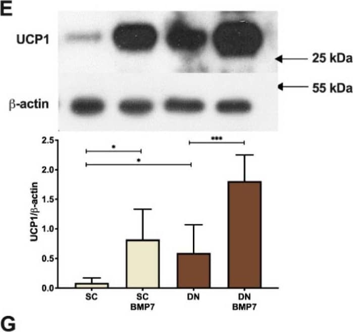
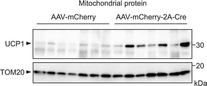
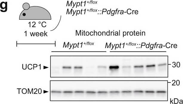
Western blot shows lysates of mouse brown adipose tissue and mouse adipose tissue. PVDF Membrane was probed with 0.5 µg/mL of Mouse Anti-Human/Mouse UCP1 Monoclonal Antibody (Catalog # MAB6158) followed by HRP-conjugated Anti-Mouse IgG Secondary Antibody (Catalog # HAF007). A specific band was detected for UCP1 at approximately 33 kDa (as indicated). This experiment was conducted under reducing conditions and using Immunoblot Buffer Group 2.UCP1 in Human Mesenchymal Stem Cells.
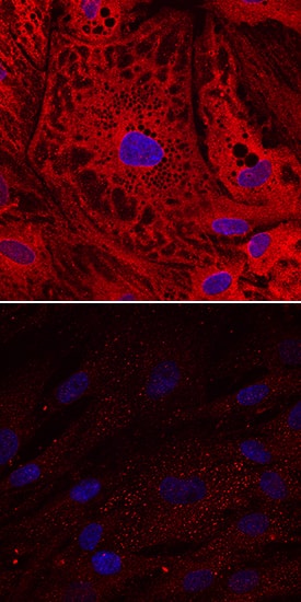
UCP1 was detected in immersion fixed human mesenchymal stem cells undifferentiated (lower panel) or differentiated into adipocytes (upper panel) using Mouse Anti-Human/Mouse UCP1 Monoclonal Antibody (Catalog # MAB6158) at 10 µg/mL for 3 hours at room temperature. Cells were stained using the NorthernLights™ 557-conjugated Anti-Mouse IgG Secondary Antibody (red; Catalog # NL007) and counterstained with DAPI (blue). Specific staining was localized to cytoplasm. View our protocol for Fluorescent ICC Staining of Stem Cells on Coverslips.Applications for Human/Mouse UCP1 Antibody
Immunocytochemistry
Sample: Immersion fixed human mesenchymal stem cells differentiated into adipocytes
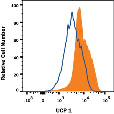
Intracellular Staining by Flow Cytometry
Sample: 3T3‑L1 mouse embryonic fibroblast adipose-like cell line fixed with Flow Cytometry Fixation Buffer (Catalog # FC004) and permeabilized with Flow Cytometry Permeabilization/Wash Buffer I (Catalog # FC005)
Simple Western
Sample: Mouse brown adipose tissue
Western Blot
Sample: Mouse brown adipose tissue and mouse adipose tissue
Reviewed Applications
Read 2 reviews rated 5 using MAB6158 in the following applications:
Formulation, Preparation, and Storage
Purification
Reconstitution
Formulation
Shipping
Stability & Storage
- 12 months from date of receipt, -20 to -70 °C as supplied.
- 1 month, 2 to 8 °C under sterile conditions after reconstitution.
- 6 months, -20 to -70 °C under sterile conditions after reconstitution.
Background: UCP1
Mitochondrial brown fat uncoupling protein 1 (UCP1; also Thermogenin and UCP) is a 33 kDa member of the mitochondrial carrier family of proteins. Human and mouse UCP1 are both 307 amino acids (aa) in length and contain three solcar repetitive regions and six transmembrane segments. UCP1 is found in brown adipose tissue, where it becomes activated by fatty acids and inhibited by nucleotides. It functions as a mitochondrial transporter that creates a proton leak across the inner mitochondrial membrane, uncoupling oxidative phosphorylation from ATP synthesis. As a result, energy is dissipated in the form of heat. Human and mouse UCP1 share 79% aa sequence identity.
Long Name
Alternate Names
Gene Symbol
UniProt
Additional UCP1 Products
Product Documents for Human/Mouse UCP1 Antibody
Product Specific Notices for Human/Mouse UCP1 Antibody
For research use only

