Human Nectin-2/CD112 Antibody
R&D Systems, part of Bio-Techne | Catalog # AF2229

Key Product Details
Validated by
Species Reactivity
Validated:
Cited:
Applications
Validated:
Cited:
Label
Antibody Source
Product Specifications
Immunogen
Gln32-Leu360
Accession # NP_002847
Specificity
Clonality
Host
Isotype
Scientific Data Images for Human Nectin-2/CD112 Antibody
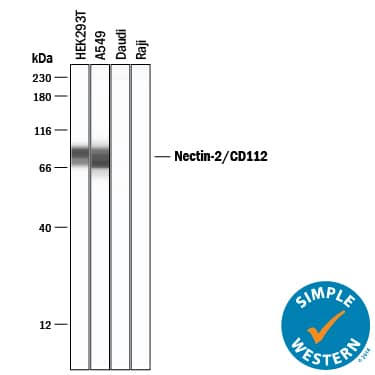
Detection of Human Nectin‑2/CD112 by Western Blot.
Western blot shows lysates of 293T human embryonic kidney cell line, A549 human lung carcinoma cell line, Daudi human Burkitt's lymphoma cell line, and Raji human Burkitt's lymphoma cell line. PVDF membrane was probed with 0.1 µg/mL of Goat Anti-Human Nectin-2/CD112 Antigen Affinity-purified Polyclonal Antibody (Catalog # AF2229) followed by HRP-conjugated Anti-Goat IgG Secondary Antibody (Catalog # HAF017). A specific band was detected for Nectin-2/CD112 at approximately 60-75 kDa (as indicated). Daudi human Burkitt's lymphoma cell line and Raji human Burkitt's lymphoma cell line are shown as negative controls. GAPDH (Catalog # AF5718) is shown as a loading control.This experiment was conducted under reducing conditions and using Immunoblot Buffer Group 1.Nectin‑2/CD112 in MCF‑7 Human Cell Line.
Nectin-2/CD112 was detected in immersion fixed MCF-7 human breast cancer cell line using Goat Anti-Human Nectin-2/CD112 Antigen Affinity-purified Polyclonal Antibody (Catalog # AF2229) at 1.7 µg/mL for 3 hours at room temperature. Cells were stained using the NorthernLights™ 557-conjugated Anti-Goat IgG Secondary Antibody (red; Catalog # NL001) and counterstained with DAPI (blue). Specific staining was localized to cytoplasm. View our protocol for Fluorescent ICC Staining of Cells on Coverslips.Nectin‑2/CD112 in Human Liver.
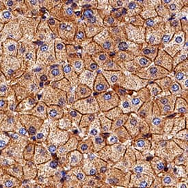
Nectin-2/CD112 was detected in immersion fixed paraffin-embedded sections of human liver using Goat Anti-Human Nectin-2/CD112 Antigen Affinity-purified Polyclonal Antibody (Catalog # AF2229) at 3 µg/mL for 1 hour at room temperature followed by incubation with the Anti-Goat IgG VisUCyte™ HRP Polymer Antibody (Catalog # VC004). Tissue was stained using DAB (brown) and counterstained with hematoxylin (blue). Specific staining was localized to cell membrane. View our protocol for IHC Staining with VisUCyte HRP Polymer Detection Reagents.Applications for Human Nectin-2/CD112 Antibody
Immunocytochemistry
Sample: Immersion fixed MCF-7 human breast cancer cell line
Immunohistochemistry
Sample: Immersion fixed paraffin-embedded sections of human liver
Simple Western
Sample: HEK293T human embryonic kidney cell line and A549 human lung carcinoma cell line
Western Blot
Sample: 293T human embryonic kidney cell line and A549 human lung carcinoma cell line
Formulation, Preparation, and Storage
Purification
Reconstitution
Formulation
Shipping
Stability & Storage
- 12 months from date of receipt, -20 to -70 °C as supplied.
- 1 month, 2 to 8 °C under sterile conditions after reconstitution.
- 6 months, -20 to -70 °C under sterile conditions after reconstitution.
Background: Nectin-2/CD112
Nectins are a small family of Ca++-independent immunoglobulin (Ig)-like cell adhesion molecules (CAMs) that organize intercellular junctions (1). The nectin family has at least four members (nectin-1-4), all of which show alternate splicing (except for Nectin-4), a transmembrane (TM) region (except for Nectin-1 gamma), and three extracellular Ig-domains. Nectins are highly homologous to the human receptor for poliovirus, and as such have been alternately named poliovirus receptor-related proteins. They do not, however, appear to bind poliovirus (1). Nectin-2 is a 60 or 65 kDa type I TM glycoprotein that is found on a variety of cell types (2, 3). It has two splice forms (4, 5). Nectin-2 delta is a 65 kDa long form and is synthesized as a 538 amino acid precursor. It contains a 31 amino acid (aa) signal sequence, a 329 aa extracellular region, a 21 aa TM segment, and a 157 aa cytoplasmic domain. The extracellular region contains one N-terminal 85 aa V-type Ig domain and two 45-55 aa C2-type Ig domains. The V-domain is believed to mediate nectin binding to its ligands (6). The short, 60 kDa isoform of Nectin-2 (Nectin-2 alpha) has the same signal sequence and extracellular domain as nectin-2 delta, but differs in the TM and cytoplasmic region (4, 5). In this case, the cytoplasmic tail is only 94 aa in length. The human extracellular region shows 72% aa sequence identity with the equivalent region in mouse. Nectin-2 is known to bind the pseudorabies virus, and herpes simplex virus-2 (HSV-2), but not HSV-1. It does not bind poliovirus. As a cell adhesion molecule, Nectin-2 will form cis-homodimers (same cell), followed by trans-dimers (across cells). Nectin-2 will not cis-dimerize with other nectins, but will cis-dimerize with its two splice forms. Notably, a Nectin-2 cis-dimer on one cell will heterodimerize with a Nectin-3 cis-dimer on another cell (1). Nectin-2 is found concentrated in adherens junctions, and exists on neurons, endothelial cells, epithelial cells and fibroblasts.
References
- Takai, Y. and H. Nakanishi, 2003, J. Cell Sci. 116:17.
- Bottino, C. et al. (2003) J. Exp. Med. 198:557.
- Pende, D. et al. (2005) Mol. Immunol. 42:463.
- Eberle, F. et al. (1995) Gene 159:267.
- Warner, M.S. et al. (1998) Virology 246:179.
- Struyf, F. et al. (2002) J. Virol. 76:12940.
Long Name
Alternate Names
Gene Symbol
UniProt
Additional Nectin-2/CD112 Products
Product Documents for Human Nectin-2/CD112 Antibody
Product Specific Notices for Human Nectin-2/CD112 Antibody
For research use only


