Human Notch-1 Intracellular Domain Antibody
R&D Systems, part of Bio-Techne | Catalog # MAB3647

Key Product Details
Species Reactivity
Applications
Label
Antibody Source
Product Specifications
Immunogen
Gly2428-Lys2556
Accession # AAG33848
Specificity
Clonality
Host
Isotype
Scientific Data Images for Human Notch-1 Intracellular Domain Antibody
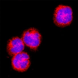
Notch-1 in MOLT-4 Human Cell Line.
Notch-1 was detected in immersion fixed MOLT-4 human acute lymphoblastic leukemia cell line using Mouse Anti-Human Notch-1 Intracellular Domain Monoclonal Antibody (Catalog # MAB3647) at 8 µg/mL for 3 hours at room temperature. Cells were stained using the NorthernLights™ 557-conjugated Anti-Mouse IgG Secondary Antibody (red; NL007) and counterstained with DAPI (blue). Specific staining was localized to cell surface and cell nuclei. Staining was performed using our protocol for Fluorescent ICC Staining of Non-adherent Cells.Applications for Human Notch-1 Intracellular Domain Antibody
Immunocytochemistry
Sample: Immersion fixed MOLT‑4 human acute lymphoblastic leukemia cell line
Western Blot
Sample: Recombinant Human Notch‑1 Intracellular Domain
Formulation, Preparation, and Storage
Purification
Reconstitution
Formulation
Shipping
Stability & Storage
- 12 months from date of receipt, -20 to -70 °C as supplied.
- 1 month, 2 to 8 °C under sterile conditions after reconstitution.
- 6 months, -20 to -70 °C under sterile conditions after reconstitution.
Background: Notch-1
Human Notch-1 is a 300 kDa type I transmembrane glycoprotein that is one of four human Notch homologues involved in developmental processes (1-3). Notch signaling is important for maintaining stem cells and inducing differentiation, especially in the nervous system and lymphoid tissues (2-4). Notch can specify binary cell fates; for example, promoting T- over B-cell development from a common precursor (2). More than 50% of human T-lineage acute lymphoblastic leukemia (T‑ALL) have activating mutations of Notch1 (1, 5). Human Notch-1 is synthesized as a 2556 amino acid (aa) precursor that contains an 18 aa signal sequence, a 1718 aa extracellular domain (ECD) with 36 EGF-like repeats and three Lin-12/Notch repeats (LNR), a 23 aa transmembrane (TM) segment and a 785 aa cytoplasmic domain containing six ankyrin repeats, a glutamine-rich domain and a PEST sequence. The 11th and 12th EGF-like repeats bind ligands including Jagged and Delta-like families in humans (6). O-fucosylation by Fringe family members at a site within this region can inhibit the interaction of Notch with Jagged ligands, thereby promoting Delta-like ligand interactions (7). Notch-1 receptor undergoes post-translational furin-type proteolytic cleavage, forming a heterodimer through interaction of a hydrophobic area C-terminal to the LNR on the 1647 aa ligand-binding extracellular region with the 891 aa transmembrane/cytoplasmic portion (8, 9). Upon ligand binding, additional sequential proteolysis by TNF-converting enzyme (ADAM17) and the presenilin-dependent gamma-secretase results in the release of the Notch intracellular domain (NICD) which translocates into the nucleus, activating transcription of Notch-responsive genes (10). Human Notch-1 ECD aa 19-526, including the first 13 EGF repeats, shows 91% aa identity with corresponding regions of mouse and rat, 89% with canine, and 79% with chicken Notch-1. This region also exhibits 60% aa identity with human Notch-2 and Notch-3.
References
- Ellisen, L.W. et al. (1991) Cell 66:649.
- Dumortier, A. et al. (2005) Int. J. Hematol. 82:277.
- Yoon, K. and N. Gaiano (2005) Nat. Neurosci. 8:709.
- Androutsellis-Theotokis, A. et al. (2006) Nature 442:823.
- Weng, A.P. et al. (2004) Science 306:269.
- Hambleton, S. et al. (2004) Structure 12:2173.
- Yang, L. et al. (2005) Mol. Biol. Cell 16:927.
- Sanchez-Irizarry, C. et al. (2004) Mol. Cell. Biol. 24:9265.
- Logeat, F. et al. (1998) Proc. Natl. Acad. Sci. USA 95:8108.
- Mumm, J.S. and R. Kopan (2000) Dev. Biol. 228:151.
Alternate Names
Gene Symbol
UniProt
Additional Notch-1 Products
Product Documents for Human Notch-1 Intracellular Domain Antibody
Product Specific Notices for Human Notch-1 Intracellular Domain Antibody
For research use only
