Human PAR2 Antibody
R&D Systems, part of Bio-Techne | Catalog # MAB3949


Key Product Details
Species Reactivity
Validated:
Cited:
Applications
Validated:
Cited:
Label
Antibody Source
Product Specifications
Immunogen
Accession # P55085
Specificity
Clonality
Host
Isotype
Scientific Data Images for Human PAR2 Antibody
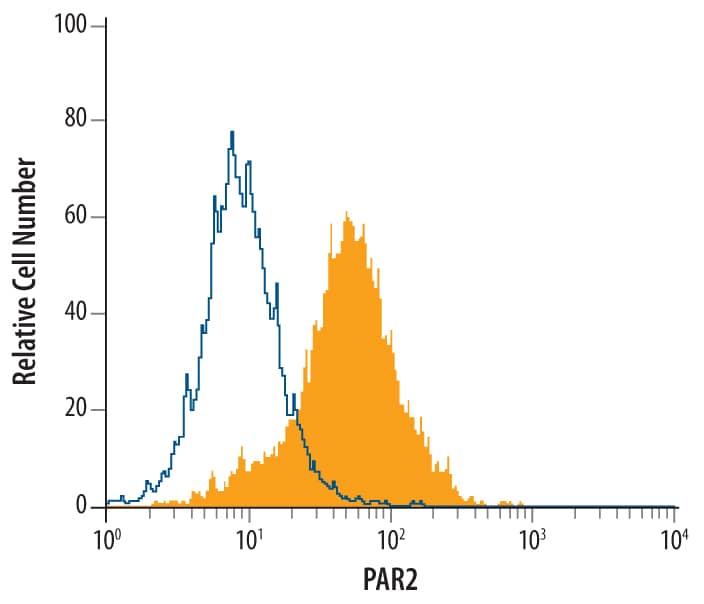
Detection of PAR2 in HT‑29 Human Cell Line by Flow Cytometry.
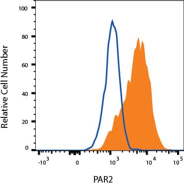
HT-29 human colon adenocarcinoma cell line was stained with Mouse Anti-Human PAR2 Monoclonal Antibody (Catalog # MAB3949, filled histogram) or isotype control antibody (MAB003, open histogram), followed by Phycoerythrin-conjugated Anti-Mouse IgG F(ab')2Secondary Antibody (F0102B).Detection of PAR2 in PC-3 cells by Flow Cytometry.
PC-3 cells were stained with Mouse Anti-Human PAR2 Monoclonal Antibody (Catalog # MAB3949, filled histogram) or isotype control antibody (Catalog # MAB003, open histogram), followed by Phycoerythrin-conjugated Anti-Mouse IgG Secondary Antibody (Catalog # F0102B). View our protocol for Staining Membrane-associated Proteins.Applications for Human PAR2 Antibody
CyTOF-ready
Flow Cytometry
Sample: HT‑29 human colon adenocarcinoma cell line and PC-3 prostate cancer cell line
Formulation, Preparation, and Storage
Purification
Reconstitution
Formulation
Shipping
Stability & Storage
- 12 months from date of receipt, -20 to -70 °C as supplied.
- 1 month, 2 to 8 °C under sterile conditions after reconstitution.
- 6 months, -20 to -70 °C under sterile conditions after reconstitution.
Background: PAR2
Protease-Activated Receptor 2 (PAR2) is a protease activated 7-transmembrane G-protein-coupled receptor. Human PAR2 contains a cleavage site for trypsin, mast cell tryptase or coagulation factor VIIa or Xa, 11 amino acids (aa) C-terminal to the signal sequence. Cleavage creates a tethered ligand that activates the 361 aa receptor. PAR2 is expressed in kidney, pancreas, stomach, intestine, airway, skin, bladder and brain; activation stimulates release of inflammatory and nociceptive mediators. PAR2 is downregulated by ubiquination, endocytosis and degradation. Mature human PAR2 shows 78% amino acid identity with mouse PAR2 over the extracellular portions.
Long Name
Alternate Names
Gene Symbol
UniProt
Additional PAR2 Products
Product Documents for Human PAR2 Antibody
Product Specific Notices for Human PAR2 Antibody
For research use only
