Human RANK/TNFRSF11A Antibody
R&D Systems, part of Bio-Techne | Catalog # MAB6832

Key Product Details
Species Reactivity
Applications
Label
Antibody Source
Product Specifications
Immunogen
extracellular domain
Accession # Q9Y6Q6
Specificity
Clonality
Host
Isotype
Scientific Data Images for Human RANK/TNFRSF11A Antibody
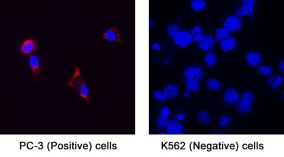
Detection of RANK/TNFRSF11A in PC-3 Human Prostate Cancer Cell Line (positive) and K562 Human Chronic Myelogenous Leukemia Cell Line (negative) Cells.
RANK/TNFRSF11A was detected in immersion fixed PC-3 Human Prostate Cancer Cell Line (positive) and K562 Human Chronic Myelogenous Leukemia Cell Line (negative) Cells using Mouse Anti-Human RANK/TNFRSF11A Monoclonal Antibody (Catalog # MAB6832) at 8 µg/mL for 3 hours at room temperature. Cells were stained using the NorthernLights™ 557-conjugated Anti-Mouse IgG Secondary Antibody (red; Catalog # NL007) and counterstained with DAPI (blue). Specific staining was localized to cytoplasm. View our protocol for Fluorescent ICC Staining of Cells on Coverslips.Detection of RANK/TNFRSF11A in Human Colon.
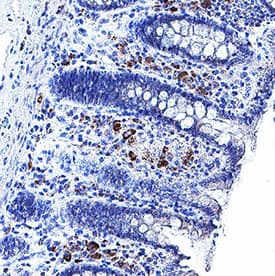
RANK/TNFRSF11A was detected in immersion fixed paraffin-embedded sections of Human Colon using Mouse Anti-Human RANK/TNFRSF11A Monoclonal Antibody (Catalog # MAB6832) at 5 µg/mL for 1 hour at room temperature followed by incubation with the Anti-Mouse IgG VisUCyte™ HRP Polymer Antibody (Catalog # VC001). Before incubation with the primary antibody, tissue was subjected to heat-induced epitope retrieval using VisUCyte Antigen Retrieval Reagent-Basic (Catalog # VCTS021). Tissue was stained using DAB (brown) and counterstained with hematoxylin (blue). Specific staining was localized to cytoplasm in lymphocytes. View our protocol for IHC Staining with VisUCyte HRP Polymer Detection Reagents.Applications for Human RANK/TNFRSF11A Antibody
Immunocytochemistry
Sample: Immersion fixed PC‑3 Human Prostate Cancer Cell Line (positive) and K562 Human Chronic Myelogenous Leukemia Cell Line (negative) Cells.
Immunohistochemistry
Sample: Immersion fixed paraffin-embedded sections of Human Colon
Formulation, Preparation, and Storage
Purification
Reconstitution
Formulation
Shipping
Stability & Storage
- 12 months from date of receipt, -20 to -70 °C as supplied.
- 1 month, 2 to 8 °C under sterile conditions after reconstitution.
- 6 months, -20 to -70 °C under sterile conditions after reconstitution.
Background: RANK/TNFRSF11A
RANK (receptor activator of NF-kappa B, also known as TRANCE receptor, osteoclast differentiation factor receptor [ODFR]) and TNFRSF11A is a member of the tumor necrosis factor receptor family. The full length human RANK cDNA encodes a type I transmembrane protein of 616 amino acids with a predicted 184 amino acid extracellular domain and a 383 amino acid cytoplasmic domain. The extracellular domain contains two potential N-linked glycosylation sites. RANK shares significant amino acid homology with other members of the TNF R family in its extracellular four cysteine-rich repeats. Human and murine RANK share 81% amino acid identity in their extracellular domains. RANK is widely expressed with highest levels in skeletal muscle, thymus, liver, colon, small intestine and adrenal gland. RANK is expressed in dendritic cells. In activated human peripheral blood T lymphocytes, RANK expression is induced by IL-4 and TGF-beta. Multiple tumor necrosis factor receptor-associated factors (TRAFs) are involved in the signaling of RANK. TRANCE (TNF-related activation-induced cytokines, also known as RANK ligand [RANKL], osteoprotegerin ligand [OPGL], and osteoclast differentiation factor [ODF]) is the ligand for RANK. The biological functions mediated through RANK include activation of NF-kappa B and c-jun N-terminal kinase, enhancement of T cell growth and dendritic cell function, induction of osteoclastogenesis, and lymph node organogenesis. Soluble RANK is able to block TRANCE induced biological activity.
References
- Anderson, D.M. et al. (1997) Nature 390:175.
- Nakagawa, N. et al. (1998) Biochem. Biophys. Res. Commun. 245:382.
Long Name
Alternate Names
Gene Symbol
UniProt
Additional RANK/TNFRSF11A Products
Product Documents for Human RANK/TNFRSF11A Antibody
Product Specific Notices for Human RANK/TNFRSF11A Antibody
For research use only

