Human VEGF Antibody
R&D Systems, part of Bio-Techne | Catalog # MAB2932


Conjugate
Catalog #
Key Product Details
Species Reactivity
Validated:
Human
Cited:
Mouse
Applications
Validated:
CyTOF-ready, Immunocytochemistry, Immunohistochemistry, Intracellular Staining by Flow Cytometry
Cited:
Mass Cytometry
Label
Unconjugated
Antibody Source
Monoclonal Mouse IgG2A Clone # VG1
Product Specifications
Immunogen
Recombinant VEGF 189 protein
Specificity
Detects human VEGF in direct ELISAs. This VEGF Antibody (Clone VG1) detects the 189, 165 and 121
isoforms of VEGF.
Clonality
Monoclonal
Host
Mouse
Isotype
IgG2A
Scientific Data Images for Human VEGF Antibody
VEGF in HeLa Human Cell Line.
VEGF was detected in immersion fixed HeLa human cervical epithelial carcinoma cell line using Mouse Anti-Human VEGF Monoclonal Antibody (Catalog # MAB2932) at 5 µg/mL for 3 hours at room temperature. Cells were stained using the NorthernLights™ 557-conjugated Anti-Mouse IgG Secondary Antibody (red; NL007) and counterstained with DAPI (blue). Specific staining was localized to cytoplasm. View our protocol for Fluorescent ICC Staining of Cells on Coverslips.VEGF in Human Liver Cancer Tissue.
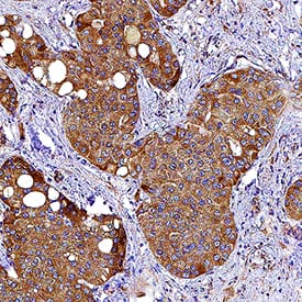
VEGF was detected in immersion fixed paraffin-embedded sections of human liver cancer tissue using Mouse Anti-Human VEGF Monoclonal Antibody (Catalog # MAB2932) at 5 µg/mL for 1 hour at room temperature followed by incubation with the Anti-Mouse IgG VisUCyte™ HRP Polymer Antibody (Catalog # VC001). Before incubation with the primary antibody, tissue was subjected to heat-induced epitope retrieval using Antigen Retrieval Reagent-Basic (CTS013). Tissue was stained using DAB (brown) and counterstained with hematoxylin (blue). Specific staining was localized to cancer cells. View our protocol for IHC Staining with VisUCyte HRP Polymer Detection Reagents.Detection of VEGF in U937 Human Cell Line by Flow Cytometry.
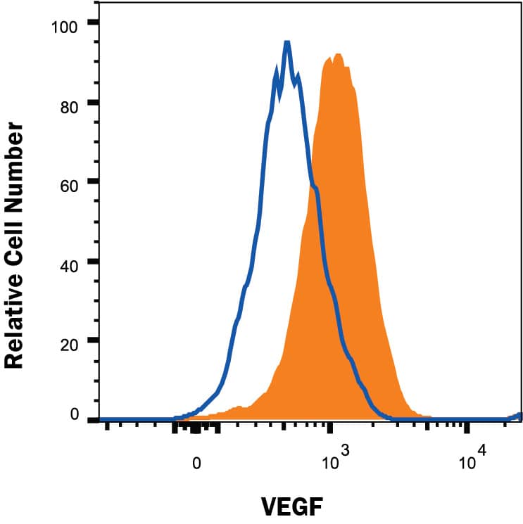
U937 human histiocytic lymphoma cell line was stained with Mouse Anti-Human VEGF Monoclonal Antibody (Catalog # MAB2932, filled histogram) or isotype control antibody (MAB002, open histogram) followed by anti-Mouse IgG APC-conjugated Secondary Antibody (F0101B). To facilitate intracellular staining, cells were fixed and permeabilized with FlowX FoxP3/Transcription Factor Fixation & Perm Buffer Kit (FC012). View our protocol for Staining Intracellular Molecules.Applications for Human VEGF Antibody
Application
Recommended Usage
CyTOF-ready
Ready to be labeled using established conjugation methods. No BSA or other carrier proteins that could interfere with conjugation.
Immunocytochemistry
5-25 µg/mL
Sample: Immersion fixed HeLa human cervical epithelial carcinoma cell line
Sample: Immersion fixed HeLa human cervical epithelial carcinoma cell line
Immunohistochemistry
5-25 µg/mL
Sample: Immersion fixed paraffin-embedded sections of human liver cancer tissue
Sample: Immersion fixed paraffin-embedded sections of human liver cancer tissue
Intracellular Staining by Flow Cytometry
0.25 µg/106 cells
Sample: U937 human histiocytic lymphoma cell line fixed with Flow Cytometry Fixation Buffer (Catalog # FC004) and permeabilized with Methanol.
Sample: U937 human histiocytic lymphoma cell line fixed with Flow Cytometry Fixation Buffer (Catalog # FC004) and permeabilized with Methanol.
Formulation, Preparation, and Storage
Purification
Protein A or G purified from hybridoma culture supernatant
Reconstitution
Reconstitute at 0.5 mg/mL in sterile PBS. For liquid material, refer to CoA for concentration.
Formulation
Lyophilized from a 0.2 μm filtered solution in PBS with Trehalose. *Small pack size (SP) is supplied either lyophilized or as a 0.2 µm filtered solution in PBS.
Shipping
Lyophilized product is shipped at ambient temperature. Liquid small pack size (-SP) is shipped with polar packs. Upon receipt, store immediately at the temperature recommended below.
Stability & Storage
Use a manual defrost freezer and avoid repeated freeze-thaw cycles.
- 12 months from date of receipt, -20 to -70 °C as supplied.
- 1 month, 2 to 8 °C under sterile conditions after reconstitution.
- 6 months, -20 to -70 °C under sterile conditions after reconstitution.
Background: VEGF
Long Name
Vascular Endothelial Growth Factor
Alternate Names
MVCD1, VAS, Vasculotropin, VEGF-A, VEGFA, VPF
Entrez Gene IDs
Gene Symbol
VEGFA
Additional VEGF Products
Product Documents for Human VEGF Antibody
Product Specific Notices for Human VEGF Antibody
For research use only
Loading...
Loading...
Loading...
Loading...
Loading...


