Mouse UTF1 Antibody
R&D Systems, part of Bio-Techne | Catalog # AF7819

Key Product Details
Validated by
Species Reactivity
Applications
Label
Antibody Source
Product Specifications
Immunogen
Lys50-Ser171
Accession # Q6J1H4
Specificity
Clonality
Host
Isotype
Scientific Data Images for Mouse UTF1 Antibody
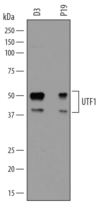
Detection of Mouse UTF1 by Western Blot.
Western blot shows lysates of D3 mouse embryonic stem cell line and P19 mouse embryonal carcinoma cell line. PVDF membrane was probed with 0.5 µg/mL of Sheep Anti-Mouse UTF1 Antigen Affinity-purified Polyclonal Antibody (Catalog # AF7819) followed by HRP-conjugated Anti-Sheep IgG Secondary Antibody (Catalog # HAF016). Specific bands were detected for UTF1 at approximately 50 and 40 kDa (as indicated). This experiment was conducted under reducing conditions and using Immunoblot Buffer Group 1.Detection of UTF1 in D3 Mouse Cell Line by Flow Cytometry.
D3 mouse embryonic stem cell line untreated (panel A) or treated with 10 mM retinoic acid for 3 days (panel B) was stained with Sheep Anti-Mouse UTF1 Antigen Affinity-purified Polyclonal Antibody (Catalog # AF7819, filled histogram) or control antibody (Catalog # 5-001-A, open histogram), followed by Phycoerythrin-conjugated Anti-Sheep IgG Secondary Antibody (Catalog # F0126). UTF1 expression is decreased with retinoic acid treatment, as indicated in panel B. To facilitate intracellular staining, cells were fixed with paraformaldehyde and permeabilized with saponin.UTF1 in D3 Mouse Stem Cells.
UTF1 was detected in immersion fixed D3 mouse embryonic stem cell line using Sheep Anti-Mouse UTF1 Antigen Affinity-purified Polyclonal Antibody (Catalog # AF7819) at 10 µg/mL for 3 hours at room temperature. Cells were stained using the Northern-Lights™ 557-conjugated Anti-Sheep IgG Secondary Antibody (red; Catalog # NL010). Cells were double-stained using Mouse Anti-Human/Mouse SSEA-1 Monoclonal Antibody (Catalog # MAB2155) and the Northern-Lights™ 493-conjugated Anti-Mouse IgG Secondary Antibody (green; Catalog # NL009). Specific staining of UTF1 was localized to nuclei. View our protocol for Fluorescent ICC Staining of Cells on Coverslips.Applications for Mouse UTF1 Antibody
CyTOF-ready
Flow Cytometry
Sample: D3 mouse embryonic stem cell line untreated or treated with retinoic acid (expression is decreased) and fixed with paraformaldehyde and permeabilized with saponin.
Immunocytochemistry
Sample: Immersion fixed D3 mouse embryonic stem cell line
Western Blot
Sample: D3 mouse embryonic stem cell line and P19 mouse embryonal carcinoma cell line
Formulation, Preparation, and Storage
Purification
Reconstitution
Formulation
Shipping
Stability & Storage
- 12 months from date of receipt, -20 to -70 °C as supplied.
- 1 month, 2 to 8 °C under sterile conditions after reconstitution.
- 6 months, -20 to -70 °C under sterile conditions after reconstitution.
Background: UTF1
UTF1 (Undifferentiated embryonic cell Transcription Factor 1) is a 47-49 kDa member of Leu-zipper family of proteins. It is expressed in the early embryonic inner cell mass and primitive ectoderm, and disappears following the formation of the primitive streak. In this regard, UTF1 has been shown to be chromatin-associated and to participate in the proper differentiation of embryonic stem cells. It also is expressed in basal keratinocytes, and hair follicle matrix cells. UTF1 acts as a transcriptional coactivator of ATF2 (Activating Transcription factor 2), a bZIP family protein that interacts with CRE in gene promoters. Mouse UTF1 is 399 amino acids (aa) in length. It contains two conserved domains (aa 52-167 and aa 269-332), the latter which is key to ATS2 activation. There is one alternative start site at Met43 that generates a 39 kDa isoform. Over aa 18-423, mouse UTF1 shares 81% and 95% aa sequence identity with human and rat UTF1, respectively.
Long Name
Alternate Names
Gene Symbol
UniProt
Additional UTF1 Products
Product Documents for Mouse UTF1 Antibody
Product Specific Notices for Mouse UTF1 Antibody
For research use only


