PAR/pADPr Antibody Best Seller
R&D Systems, part of Bio-Techne | Catalog # 4335-MC-100

Key Product Details
Species Reactivity
Validated:
Multi-Species
Cited:
Human, Mouse, Rat, N/A, Transgenic Mouse
Applications
Validated:
Immunocytochemistry, Immunohistochemistry, Intracellular Staining by Flow Cytometry, Western Blot
Cited:
Chromatin Immunoprecipitation (ChIP), Co-Immunoprecipitation, ELISA Capture, Flow Cytometry, Functional Assay, Immunocytochemistry, Immunohistochemistry, Immunohistochemistry-Paraffin, Immunoprecipitation, Proximity Ligation Assay, Westen Blot, Western Blot, Western Blot Control
Label
Unconjugated
Antibody Source
Monoclonal Mouse IgG3 Clone # 10HA
Product Specifications
Immunogen
Purified ADP-ribose polymers between 2 and 50 units long
Specificity
The antibody is specific for PAR polymers 2 to 50 units long, but does not recognize structurally related RNA, DNA, ADP-ribose monomers, NAD, or other nucleic acid monomers. Detects Poly(ADP-ribose) Polymer in Direct ELISA.
Clonality
Monoclonal
Host
Mouse
Isotype
IgG3
Scientific Data Images for PAR/pADPr Antibody
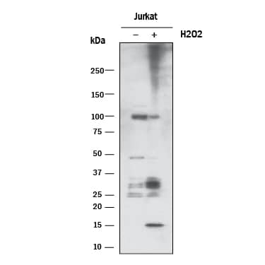
Detection of Human PAR/pADPr by Western Blot.
Western blot shows lysates of Jurkat human acute T cell leukemia cell line untreated (-) or treated (+) with 2 mM H2O2 for 5 minutes. PVDF membrane was probed with 1:1000 dilution of Mouse Anti-PAR/pADPr Monoclonal Antibody (Catalog # 4335-MC-100) followed by HRP-conjugated Anti-Mouse IgG Secondary Antibody (HAF018). Specific bands were detected for PAR polymers, as indicated. This experiment was conducted under reducing conditions and using Western Blot Buffer Group 1.Detection of PAR in Jurkat cells by Flow Cytometry.
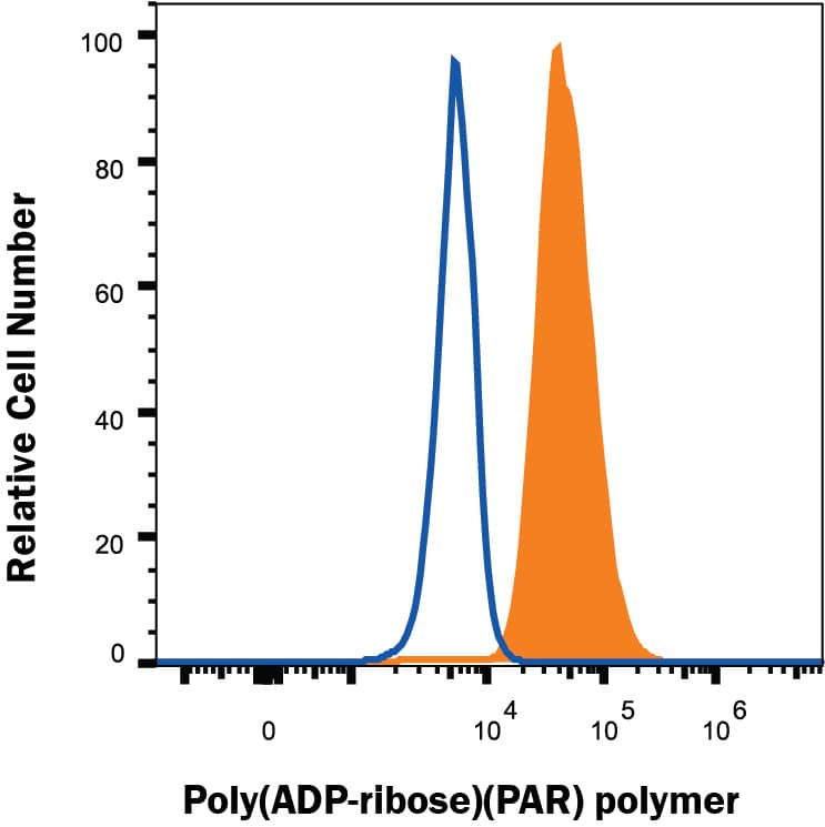
Jurkat human acute T cell leukemia cell line treated with 1 mM H2O2 for 5 minutes (filled histogram) or resting (open histogram) were stained with Mouse Anti-PAR Monoclonal Antibody (Catalog # 4335-MC-100) followed by Goat anti-Mouse IgG PE-conjugated Secondary Antibody (F0102B). To facilitate intracellular staining, cells were fixed and permeabilized using FlowX FoxP3/Transcription Factor Fixation & Perm Buffer Kit (FC012). Staining was performed using our Staining Intracellular Molecules protocol.Detection of PAR/pADPr in Human Kidney.
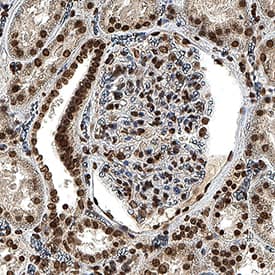
PAR/pADPr was detected in immersion fixed paraffin-embedded sections of human kidney using Mouse Anti-PAR/pADPr Monoclonal Antibody (Catalog # 4335-MC-100) at 1.7 µg/ml for 1 hour at room temperature followed by incubation with the Anti-Mouse IgG VisUCyte™ HRP Polymer Antibody (Catalog # VC001). Before incubation with the primary antibody, tissue was subjected to heat-induced epitope retrieval using VisUCyte Antigen Retrieval Reagent-Basic (Catalog # VCTS021). Tissue was stained using DAB (brown) and counterstained with hematoxylin (blue). Specific staining was localized to the nucleus. View our protocol for IHC Staining with VisUCyte HRP Polymer Detection Reagents.Applications for PAR/pADPr Antibody
Application
Recommended Usage
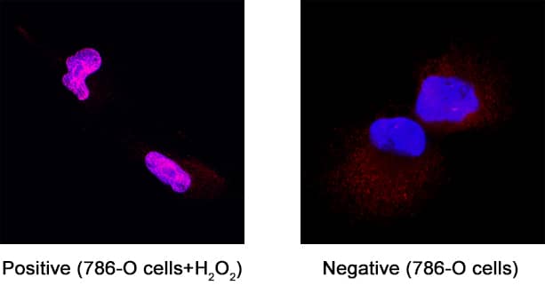
Immunocytochemistry
3-25 µg/mL
Sample: Immersion fixed 786-O human renal cell adenocarcinoma cell line
Sample: Immersion fixed 786-O human renal cell adenocarcinoma cell line
Immunohistochemistry
1-15 µg/mL
Sample: Immersion fixed paraffin-embedded sections of human kidney
Sample: Immersion fixed paraffin-embedded sections of human kidney
Intracellular Staining by Flow Cytometry
0.25 µg/106 cells
Sample: Jurkat human acute T cell leukemia cell line treated with 1 mM H2O2 for 5 minutes
Sample: Jurkat human acute T cell leukemia cell line treated with 1 mM H2O2 for 5 minutes
Western Blot
1:1000 dilution
Sample: Jurkat human acute T cell leukemia cell line treated with H2O2
Sample: Jurkat human acute T cell leukemia cell line treated with H2O2
Reviewed Applications
Read 3 reviews rated 4 using 4335-MC-100 in the following applications:
Formulation, Preparation, and Storage
Purification
Protein A or G purified from ascites
Formulation
Supplied as 100 μL of a 0.2
μm filtered solution in TBS, at a concentration of 1 mg/mL
Shipping
The product is shipped with dry ice or equivalent. Upon receipt, store it immediately at the temperature recommended below.
Stability & Storage
Use a manual defrost freezer and avoid repeated freeze-thaw cycles.
- 12 months from date of receipt, -20 to -70 °C, as supplied.
- 1 month, 2 to 8 °C under sterile conditions after opening.
- 6 months, -20 to -70 °C under sterile conditions after opening.
Background: PAR/pADPr
Long Name
Poly [ADP-ribose] Polymer
Alternate Names
PAR
Additional PAR/pADPr Products
Product Documents for PAR/pADPr Antibody
Product Specific Notices for PAR/pADPr Antibody
For research use only
Loading...
Loading...
Loading...
Loading...
Loading...


